Category: Zonder categorie

Anterior pelvic tilt assessment

VOETBAL MEDISCH SYMPOSIUM 2020

DE BEHANDELING VAN VOETBALBLESSURES 

PRAKTISCHE WETENSCHAP

OP DE KNVB CAMPUS IN ZEIST VINDT KOMEND JAAR OPNIEUW HET VOETBALMEDISCH SYMPOSIUM PLAATS.

HET SYMPOSIUM IS DÉ PLEK OM COLLEGA’S BINNEN HET VOETBALMEDISCHE DOMEIN TE ONTMOETEN OF KENNIS OP TE DOEN VAN GERENOMMEERDE EXPERTS. EN DIE NIEUWSTE INNOVATIES TE ZIEN OP HET GEBIED VAN VOETBALMEDISCHE EN FYSIEKE PRESTATIES.

NA VORIG JAAR DE DIAGNOSTIEK VAN VOETBALBLESSURES BELICHT TE HEBBEN, ROLT DE BAL DIT JAAR VERDER NAAR DE BEHANDELING VAN VOETBALBLESSURES. HET INHOUDELIJKE PROGRAMMA BIEDT OPNIEUW SPREKERS DIE ZICH ONDERSCHEIDEN IN ZOWEL DE DAGELIJKSE ZORG VOOR DE VOETBALLERS ALS OP WETENSCHAPPELIJK GEBIED.

Ja, ik wil mij aanmelden!

VOETBAL MEDISCHE WORKSHOP 2020

(VELD)REVALIDATIE NA EEN VOETBALBLESSURE

OP 4 MAART ZAL ER WEDEROM EEN WORKSHOP PLAATS VINDEN BIJ HET KNVB VOETBAL MEDISCH CENTRUM. 
OOK DIT JAAR BELOOFD HET EEN OCHTENDVULLEND PROGRAMMA TE ZIJN WAAR VOORNAMELIJK (SPORT)FYSIOTHERAPEUTEN HUN KENNIS MEE KUNNEN UITBREIDEN.

TIJDENS DE WORKSHOP ZAL MATT TABERNER ZIJN KENNIS EN EXPERTICE MET DE DEELNEMERS GAAN DELEN. MATT TABERNER IS EEN ERVAREN CLINICUS DIE AL JAREN EINDVERANTWOORDELIJK IS VOOR DE REVALIDATIE VAN TOPVOETBALLERS IN DE PREMIER LEAGUE. ZIJN FOCUS LIGT VOORNAMELIJK OP FYSIEKE ONTWIKKELING EN PRESTATIES. TEVENS IS HIJ DE ONTWIKKELAAR VAN HET ‘CONTROL-CHAOS CONTINUUM’.

DIT FRAMEWORK, WELKE VIJF FASES BESCHRIJFT HOE DE VELDREVALIDATIE NA EEN VOETBALBLESSURE OPGEBOUWD KAN WORDEN, STAAT CENTRAAL BINNEN DE WORKSHOP. DE THEORETISCHE ACHTERGROND,
DE TOEPASSING EN HET PRAKTISCHE ASPECT ZULLEN ALLEN AAN BOD KOMEN TIJDENS DE WORKSHOP.

JA, IK WIL MIJ AANMELDEN!

reviews

These guys are the best when it comes to style and have great attention to detail. 

Issac Newton
MODEL, FRANCE

These guys are the best when it comes to style and have great attention to detail. 

Issac Newton
MODEL, FRANCE

These guys are the best when it comes to style and have great attention to detail. 

Issac Newton
MODEL, FRANCE

Nick van der Horst

Meet the soccerdoc

Nick van der Horst behaalde zijn diploma fysiotherapie in 2007 aan de Hogeschool Utrecht. Hij werkte 10 jaar lang als sportfysiotherapeut/echografist/docent bij het Academie Instituut te Utrecht. Daarna heeft hij de overstap gemaakt naar waar zijn hart ligt, het professionele voetbal. Hij heeft twee jaar als sportfysiotherapeut en hoofd van de medische staf bij Go Ahead Eagles in Deventer gewerkt. Momenteel is is Nick werkzaam bij de KNVB. Zijn onderzoeks-activiteiten zijn gefocust op de voetbal-medische zorg. In 2017 behaalde hij zijn doctoraal na het verdedigen van zijn proefschrift ‘Prevention of hamstring injuries in male soccer’.

Blogger: Raúl Gómez
Blogger: Raúl Gómez

Correct pelvic alignment – ​​Posture assessment
Although there is controversy regarding the analysis of static posture and its relationship with injuries during sports movements, certain postural patterns can provide valuable information about certain players’ predispositions to injury.

Recent studies have highlighted the role of excessive anterior pelvic tilt (APT) in increasing the risk of hamstring (Mendiguchia, et al., 2021) and hip injuries (Suits, 2021) and low back pain (Das, et al., 2017). However, the field is still evolving, and there are no cut-off values for the degree of pelvic tilt and its relationship to the most common injuries in football.

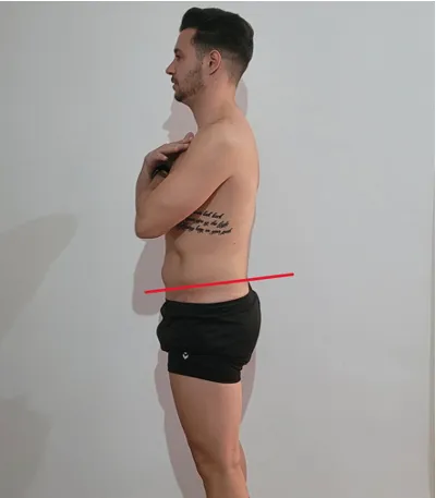
Suits (2021) defined pelvic tilt as the angle formed by a horizontal line and a line that separates the anterior superior iliac spine and the posterior superior iliac spine in the sagittal plane (see Image 1). He also presented in his article the findings of several articles that measured the pelvic tilt angle in healthy subjects, showing results ranging from 8º to 13º APT. This indicates that APT is a natural anatomical position and not inherently harmful. However, problems can arise when excessive APT occurs, particularly during high-intensity running.
SoccerDoc Blogs EU

Image 1. Pelvic tilt

Falk et al. (2020) set a cut-off value of 8° for APT, considering a higher degree excessive. However, the previous review by Suits (2021) suggests that this
cut-off value may be too low. Additionally, in healthy populations, women may exhibit a higher degree of APT than men (11.7° vs. 9.6°) (McKeon & Hertel, 2009).

In a 2013 study, Barbosa et al. compared the impact of a treatment involving manipulation of the sacroiliac joint and strength training of knee flexors and extensors on the degree of APT and subjective pain in 7 patients with low back pain. The results demonstrated a reduction of more than 5 degrees in APT (from 20.38 degrees to 14.63 degrees), which also led to a significant decrease in pain as indicated by subjective pain scale measurements (from 5.83 to 1.29).

Can we classify an APT greater than 13 degrees as a faulty postural pattern that increases the risk of injury in soccer players?
We can only make assumptions at this point, as no articles have studied the degree of APT and its association with injuries in football.

Range of motion

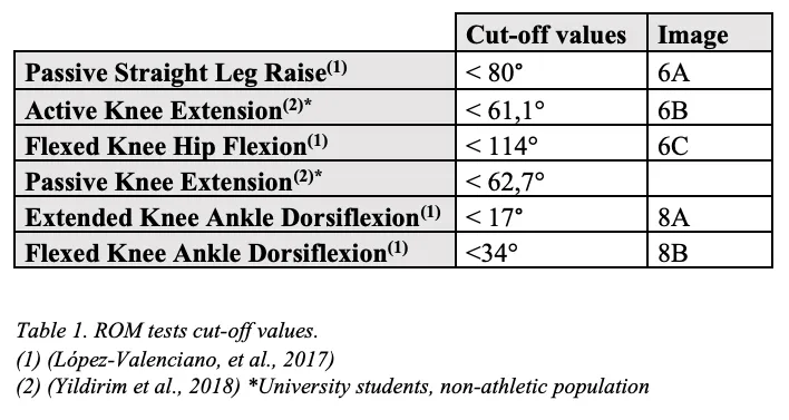
If the hip flexors are excessively tight and not adequately stabilized, they can cause the pelvis to rotate forward (Neumann, 2010). Table 1 lists the muscles that can potentially function as hip flexors and consequently increase APT.

SoccerDoc Blog EU Pelvic Tilt

Table 1. Musculature involved in hip flexion (Neumann, 2010)

Modified Thomas Test

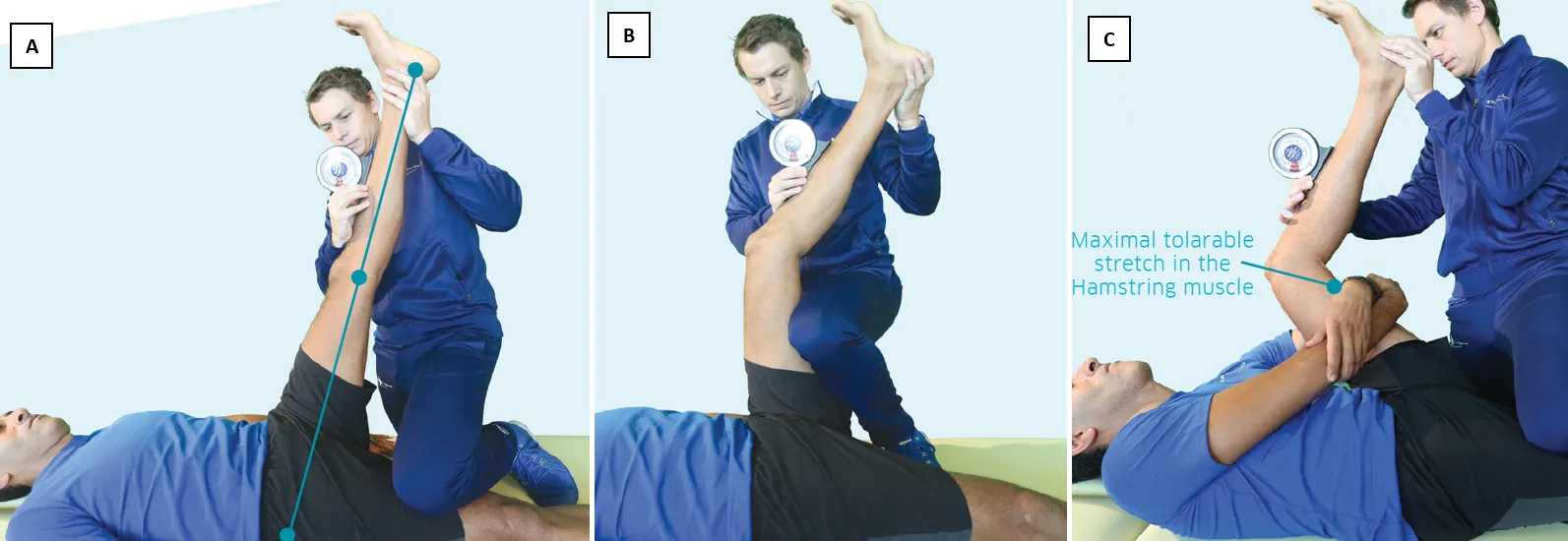
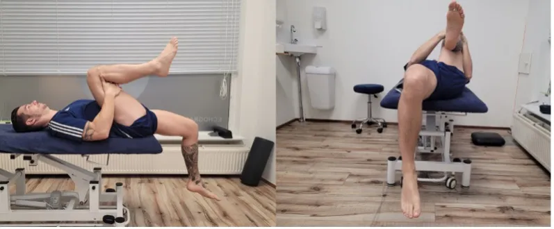
The procedure starts with the individual sitting at the edge of the table and lying back. Then, they should flex both hips as much as possible and hold their legs behind the knees in the popliteal area. Finally, they need to relax and extend one leg while still holding the other firmly in the fully flexed position (see Image 2).
SoccerDoc Blog Pelvic Tilt

Image 2. Modified Thomas Test

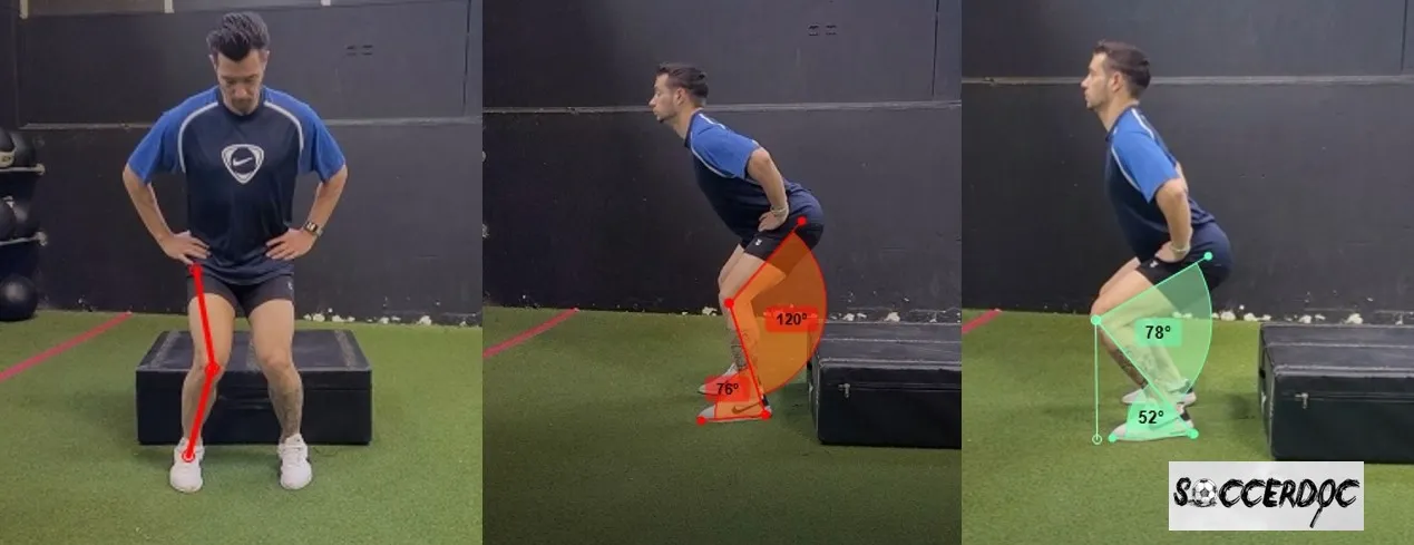
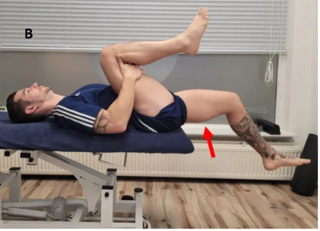
The inability to maintain a neutral back position, arching the lower back, often indicates excessive shortening of the Psoas major.
This shortening can also result in an inability to fully extend the hip (Image 3).
SoccerDoc Blog Pelvic Tilt

Image 3. Hip extension restriction

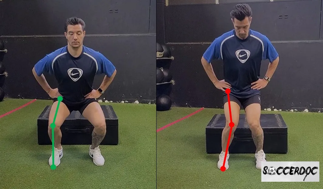
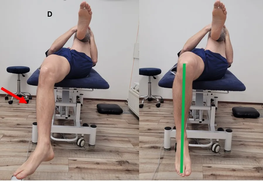
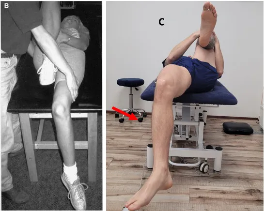
If the inability to fully extend the hip is accompanied by external rotation of the knee (Image 4, right), our biggest suspect will be the TFL. We can check this by passively adducting the extended leg (Image 4, left). An increase in external rotation of the knee will indicate excessive tension of the TFL and the iliotibial band (Neumann, 2010).
SoccerDoc Blog Pelvic Tilt

Image 4. Modified Thomas Test – Knee external rotation. (Sahrmann, 2002)

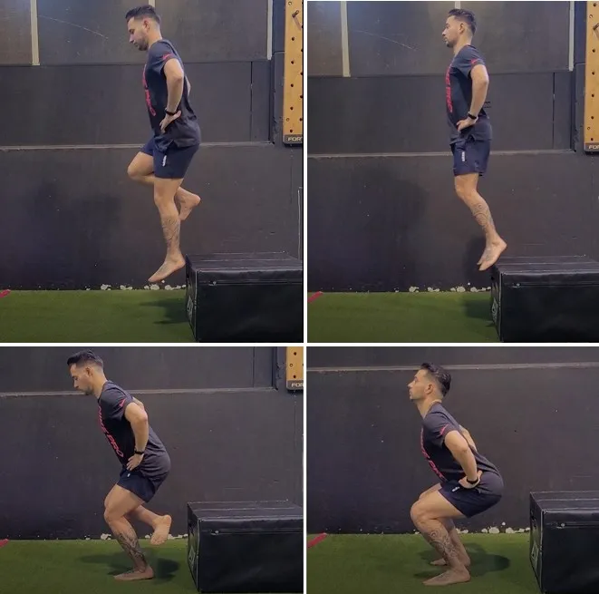
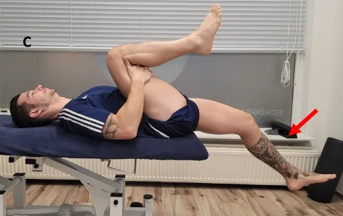
Another very common pattern is a lack of knee flexion (Image 5). Due to its biarticular nature, the rectus femoris can limit hip extension and knee flexion. Excessive tension of this muscle will cause the knee to extend, which, with correct mobility, should be perpendicular to the ground (Image 2).
SoccerDoc Blog Pelvic Tilt

Image 5. Knee extension restriction

Jurdan Test

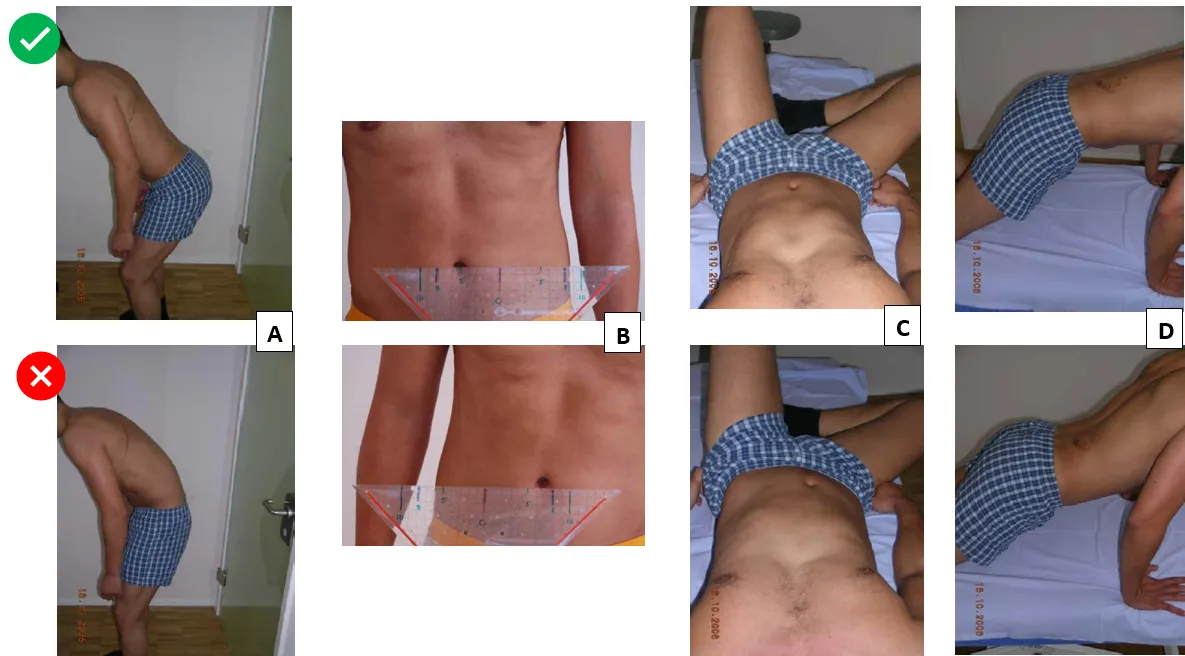
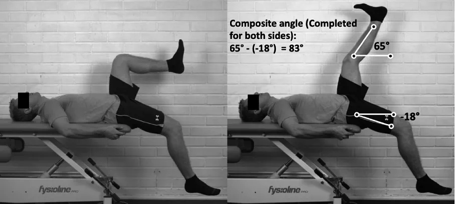
This test was first used in injury risk research in 2022 by Lahti et al. The test aims to assess the influence of the muscles of the lumbopelvic region on hamstring flexibility. The Jurdan test can be considered a combination of the active knee extension test and the modified Thomas test (Lahti, et al., 2022). The result of this test is defined as the difference between the shin angle of the actively lengthened leg and the thigh angle of the opposite leg (Image 6).
SoccerDoc Blog Pelvic Tilt

Image 6. Jurdan Test (Lahti, et al., 2022)

Although both the modified Thomas Test and the Jurdan test can provide a lot of information about hip range of motion, more research is still needed to determine cutoff values and their relation with lower limb injuries.

Strength
The abdominal muscles must generate a strong stabilizing force to counteract the strong APT caused by the hip flexors. This stabilizes the lumboabdominal region by increasing intra-abdominal pressure and thoracolumbar fascia tension (Neumann, 2010).

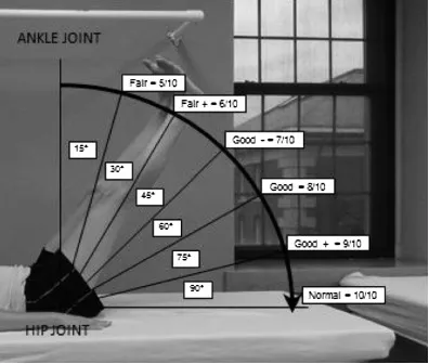
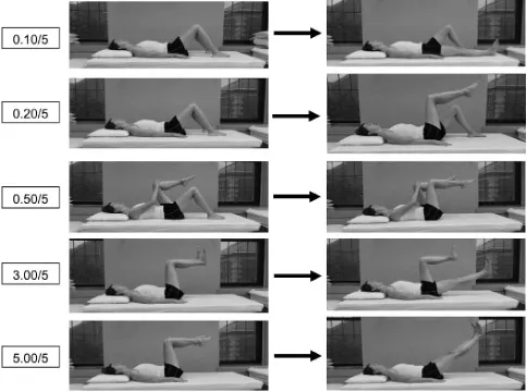
To assess the strength of the abdominal musculature against APT, Haladay et al. (2014) studied the results of the Double Limb Lowering Test* (DLLT) (Image 7) and Lower Abdominal Muscle Progression* (LAMP) (Image 8) after an abdominal stabilization exercise program. They found that the LAMP test is more sensitive to change after abdominal stabilization programs. However, no subject was able to reach the “normal” range of motion in the DLLT test as it was found to be too demanding. It is important to note that this study had some limitations, including a small number of participants (only 11), the inclusion of subjects of different genders, and varying levels of physical activity. In sports with physically demanding requirements such as football, athletes should be able to perform either of these tests with maximum results.
SoccerDoc Blog Pelvic Tilt

Image 7. DLLT. The subject lays supine and raises their legs to a position of 90º of hip flexion with the knees extended. They are then instructed to maintain their pelvis in a posterior pelvic tilt by contracting the abdominals while slowly lowering the legs to the supporting surface. Strength is graded, from 5/10 to 10/10, by identifying the point at which the subject was last able to keep the pelvis stable, as assessed by an anterior tilt of the pelvis and arching of the back (Haladay, et al., 2014).
SoccerDoc Blog Pelvic Tilt

Image 8. The subject lays supine and performs a series of 9 progressively more challenging lower extremity combinations of movements, assessing their ability to stabilize the pelvis. During this test, the subject is instructed to contract their abdominal muscles by pulling their navel toward their spine. They are assigned a grade (0.10 through 5.00/5.00) for the most challenging lower extremity movement they are able to complete without motion on their pelvis or lower back (Haladay, et al., 2014).

* More information about these tests can be found in the following references: (Haladay, et al., 2014); (Sahrmann, 2002)

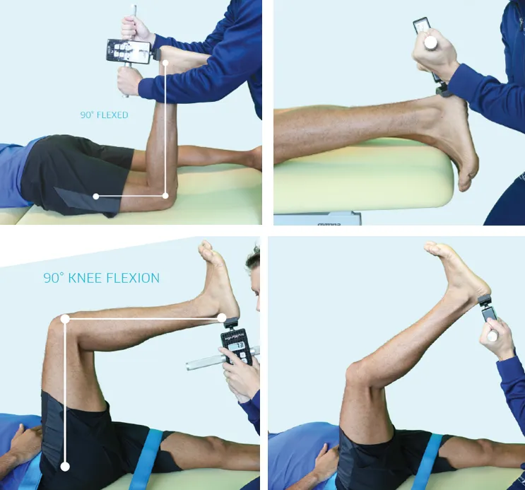
In a previous blog, we discussed the importance of the gluteus maximus (GM) muscle in correcting APT. According to Buckthorpe et al. (2019), there are various ways to assess GM strength. They affirm the prone hip extension task with the knee flexed at 90º as the most commonly used assessment and suggest using a dynamometer to measure it. Other assessment methods include isometric or isokinetic assessment. Electromyography is the most commonly used method to measure muscle activation.

Although all of these methods provide objective and reliable data, many teams may not have the necessary equipment or time to perform them.
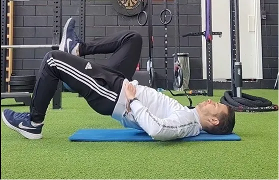
The Single-Leg Glute Bridge Test (Image 9) offers an alternative that requires no equipment and can be done with the entire team at the same time in just a few minutes.
SoccerDoc Blog Pelvic tilt

Image 9. Single-leg glute bridge

During this test, footballers are asked to perform as many repetitions as possible in the single-leg glute bridge exercise. After the test, the following data is recorded:

Maximum number of repetitions per leg – This will indicate whether there is an imbalance in the strength of the lower limb in unilateral hip extension.

Muscle that has suffered the greatest fatigue during the test – This test will determine if the muscle activation pattern of hip extension is functionally efficient. At the end of the test, it is normal to feel that fatigue in the GM has been the limiting factor of the exercise. I can assure you that you will strongly feel a “burning” sensation in that muscle if the test is carried out to failure. If other muscles, such as the quadriceps or hamstrings, are limiting the test result, this will indicate that the GM is not functioning correctly, and other muscles may be compensating for its weakness, known as synergistic dominance.

Muscle cramps or pain that may have limited the test result – Cramps in the hamstrings or back pain may indicate again weakness in the gluteus maximus and provide insight into other overloaded muscle groups due to lack of glute activation.

Motor control

Hip flexion-extension pattern

This is especially important in young players. Correctly teaching lumbopelvic dissociation is crucial, assuring that the footballer has perfect control over the motion of his/her hip without any compensation at the lumbar level. In a previous blog post, “Hamstring injuries – Post injury evaluation”, you can find several examples for the evaluation of lumbopelvic motor control.

Link: Hamstring injuries – Post injury assessment

Sprint technique

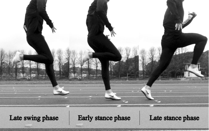
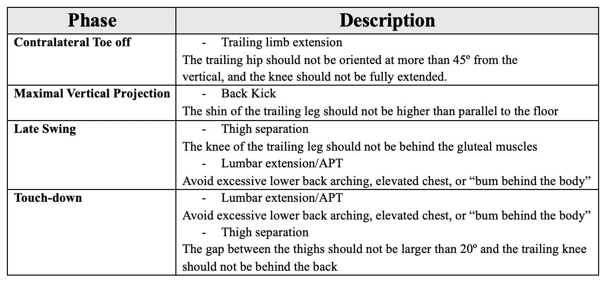
Excessive APT during sprinting increases the risk of hamstring injuries. Correcting this movement pattern is crucial. This year, the reliability of the Sprint Mechanics Assessment Score (S-MAS) was evaluated, showing it to be a reliable tool for assessing sprint mechanics in football players (Bramah et al., 2024). I recommend reading the full assessment. In this post, we will only describe the most important points for footballers who present excessive APT.

SoccerDoc Blog Pelvic Tilt

Image 10. Phases of sprint cycle. MVP – Maximal vertical projection (Bramah, et al., 2024)
SoccerDoc Blog Pelvic Tilt

Table 2. Phases of sprint cycle (Bramah, et al., 2024)

Although injury prevention is multifactorial in nature, correct and efficient movement plays a key role. Human movement is highly complex, but mastering specific motor patterns can substantially lower the risk of injury. We will soon delve into training these patterns and achieving a proper balance of muscular forces in the lumbopelvic area.

References

Barbosa, A., Martins, F., Barbosa, M. & Dos Santos, R., 2013. Manipulation and selective exercises decrease pelvic anteversion and low-back pain: A pilot study. Journal of Back and Musculoskeletal Rehabilitation, Volumen 26, pp. 33-36.

Bramah, C. y otros, 2024. The Sprint Mechanics Assessment Score – A Qualitative Screening Tool for the In-field Assessment of Sprint Running Mechanics. The American Journal of Sports Medicine, 20(10), pp. 1-9.

Buckthorpe, M., Stride, M. & Della Villa, F., 2019. Assessing and treating gluteus maximus weakness – A clinical commentary. The International Journal of Sports Physical Therapy, pp. 655-670.

Das, S. y otros, 2017. Prevalence of lower crossed syndrome in young adults: a cross sectional study. International Journal of Advanced Research, 5(6), pp. 2217-2228.

Falk, A., Overgaard, S., Hróbjartsson, A.  Holsgaard, A., 2020. Non-surgical interventions for excessive anterior pelvic tilt in synptomatic and non-symptomatic adults: A systematic review. Efort Open Reviews, Volumen 5.

Haladay, D., Miller, S., Challis, J. & Denegar, C., 2014. Responsiveness of the double limb lowering test and lower abdominal muscle progression to core stabilization exercise programs in healthy adults: A pilot study. The Journal of Strength and Conditioning Research, 28(7), pp. 1920-1927.

Lahti, J., Mendiguchia, J., Edouard, P. & Morin, J.-B., 2022. A novel multifactorial hamstring screening protocol: association with hamstring muscle injuries in professional football (soccer) – a prospective cohort study. Biology of sport, 39(4), pp. 1021-2031.

McKeon, J. Hertel, J., 2009. Sex differences and representative values for six lower extremity alignment measures. Journal of Athletic Training, 44(3), pp. 249-255.

Mendiguchia, J. y otros, 2021. Can we modify maximal speed running posture? Implications for performance and hamstring injury management. International Journal of Sports Physiology and Performance.

Neumann, D., 2010. Kinesiology of the hip: A focus on muscular actions. Journal of orthopaedic & sports physical therapy, 40(2), pp. 82-94.

Neumann, D., 2010. Kinesiology of the musculoskeletal system. 2nd ed. Missouri: Mosby.

Sahrmann, S., 2002. Diagnosis and Treatment of Movement Impairment Syndromes. 1st ed. Missouri: Mosby.

Task design in football and training load

VOETBAL MEDISCH SYMPOSIUM 2020

DE BEHANDELING VAN VOETBALBLESSURES 

PRAKTISCHE WETENSCHAP

OP DE KNVB CAMPUS IN ZEIST VINDT KOMEND JAAR OPNIEUW HET VOETBALMEDISCH SYMPOSIUM PLAATS.

HET SYMPOSIUM IS DÉ PLEK OM COLLEGA’S BINNEN HET VOETBALMEDISCHE DOMEIN TE ONTMOETEN OF KENNIS OP TE DOEN VAN GERENOMMEERDE EXPERTS. EN DIE NIEUWSTE INNOVATIES TE ZIEN OP HET GEBIED VAN VOETBALMEDISCHE EN FYSIEKE PRESTATIES.

NA VORIG JAAR DE DIAGNOSTIEK VAN VOETBALBLESSURES BELICHT TE HEBBEN, ROLT DE BAL DIT JAAR VERDER NAAR DE BEHANDELING VAN VOETBALBLESSURES. HET INHOUDELIJKE PROGRAMMA BIEDT OPNIEUW SPREKERS DIE ZICH ONDERSCHEIDEN IN ZOWEL DE DAGELIJKSE ZORG VOOR DE VOETBALLERS ALS OP WETENSCHAPPELIJK GEBIED.

Ja, ik wil mij aanmelden!

VOETBAL MEDISCHE WORKSHOP 2020

(VELD)REVALIDATIE NA EEN VOETBALBLESSURE

OP 4 MAART ZAL ER WEDEROM EEN WORKSHOP PLAATS VINDEN BIJ HET KNVB VOETBAL MEDISCH CENTRUM. 
OOK DIT JAAR BELOOFD HET EEN OCHTENDVULLEND PROGRAMMA TE ZIJN WAAR VOORNAMELIJK (SPORT)FYSIOTHERAPEUTEN HUN KENNIS MEE KUNNEN UITBREIDEN.

TIJDENS DE WORKSHOP ZAL MATT TABERNER ZIJN KENNIS EN EXPERTICE MET DE DEELNEMERS GAAN DELEN. MATT TABERNER IS EEN ERVAREN CLINICUS DIE AL JAREN EINDVERANTWOORDELIJK IS VOOR DE REVALIDATIE VAN TOPVOETBALLERS IN DE PREMIER LEAGUE. ZIJN FOCUS LIGT VOORNAMELIJK OP FYSIEKE ONTWIKKELING EN PRESTATIES. TEVENS IS HIJ DE ONTWIKKELAAR VAN HET ‘CONTROL-CHAOS CONTINUUM’.

DIT FRAMEWORK, WELKE VIJF FASES BESCHRIJFT HOE DE VELDREVALIDATIE NA EEN VOETBALBLESSURE OPGEBOUWD KAN WORDEN, STAAT CENTRAAL BINNEN DE WORKSHOP. DE THEORETISCHE ACHTERGROND,
DE TOEPASSING EN HET PRAKTISCHE ASPECT ZULLEN ALLEN AAN BOD KOMEN TIJDENS DE WORKSHOP.

JA, IK WIL MIJ AANMELDEN!

reviews

These guys are the best when it comes to style and have great attention to detail. 

Issac Newton
MODEL, FRANCE

These guys are the best when it comes to style and have great attention to detail. 

Issac Newton
MODEL, FRANCE

These guys are the best when it comes to style and have great attention to detail. 

Issac Newton
MODEL, FRANCE

Nick van der Horst

Meet the soccerdoc

Nick van der Horst behaalde zijn diploma fysiotherapie in 2007 aan de Hogeschool Utrecht. Hij werkte 10 jaar lang als sportfysiotherapeut/echografist/docent bij het Academie Instituut te Utrecht. Daarna heeft hij de overstap gemaakt naar waar zijn hart ligt, het professionele voetbal. Hij heeft twee jaar als sportfysiotherapeut en hoofd van de medische staf bij Go Ahead Eagles in Deventer gewerkt. Momenteel is is Nick werkzaam bij de KNVB. Zijn onderzoeks-activiteiten zijn gefocust op de voetbal-medische zorg. In 2017 behaalde hij zijn doctoraal na het verdedigen van zijn proefschrift ‘Prevention of hamstring injuries in male soccer’.

Blogger: Raúl Gómez
Blogger: Raúl Gómez

“These players are not fit. They need to run more during training.”  How many times have we heard this after witnessing a soccer team lose a game? Although soccer is very physically demanding, the idea that running more solves all problems is far from the truth. Football primarily depends on technical and tactical skills, with decision-making playing a crucial role.

I believe that the primary goal of training is to enhance the team’s game model, which encompasses tactical, technical, and physical aspects that are inseparable due to the nature of the game. This involves developing collective behaviors that enable the team to surpass the opposition when attacking and withstand them when defending.

Is it possible to achieve comparable improvements through training based on the team’s game model instead of isolated running training?

Bucheit et al. (2024), in their article “The 11 evidence-informed and inferred principles of microcycle periodization in football”, state the following:

“While metrics such as High-Speed Running distance and maximal speed exposures provide insights into performance and injury risks, they should not detract from the primary goal of football training: to enhance player interactions and the development of dynamic in-game scenarios through ball play.[…] It advocates for the crafting of training sessions that organically incorporate locomotor “targets” within a football-specific framework (as running is an integral part of football by definition), eliminating the necessity for isolated running drills.”

The belief that the game itself is not a valid tool to improve footballers’ physical capacity is a misconception inherited from traditional forms of periodization in other disciplines. Although certain forms of training contribute to maximizing footballers’ performance, we must not lose sight of the essence of the game.

In 2019, Kunz et al. compared the effects of high-intensity interval training (HIIT) with those of Small-Sided Games (SSG) on physical parameters in young soccer players. They concluded that SSG and HIIT result in similar improvements in several variables related to running performance. This conclusion aligns with the results of a systematic review by Hill-Haas et al. in 2011, supporting this notion.

The results regarding the improvement of neuromuscular parameters are contradictory. Some authors, such as Kunz et al. (2019) and Clemente et al. (2021), did not find effective SSGs for improving RSA, sprint, or jump. 

However, others, such as Eniseler et al. (2017), did show significant improvements in RSA after an SSG-based training program. The variability of the game, the heterogeneity in the study populations, and the differences in the design of the training programs could be the causes of these differences. With an adequate design, improvements in these physical qualities can also be achieved.

Maximum speed training with SSG seems more complex due to the unpredictable and uncontrolled nature of the game. This can lead to some soccer players overtraining while others may undertrain (Gualtieri et al., 2023).

It’s important to remember that the amount of time a player spends with the ball is crucial. In a study by Eniseler et al. (2017), one group of young football players focused their training on small-sided games (SSG), while the other group trained with linear sprints and changes of direction.

The SSG group completed a total of 5111 actions with the ball, while the other group DID NOT PERFORM ANY actions with the ball during that part of the training. As a result, the SSG group showed twice as much improvement in a short-pass test compared to the other group.

When appropriately designed, SSG are an effective tool for enhancing cardiovascular and neuromuscular fitness in football players while improving their technical and tactical skills specific to the team’s game model. However, it is important to note that other alternative methods may be more effective for improving maximum speed actions (Zhi Yong et al., 2023), as they offer simpler control and quantification.

How do different variables in task design impact the internal and external training load?

Pitch size and number of players are the variables that have the most significant impact on the physical, physiological, and technical-tactical demands (Castellano & Casamichana, 2019).

Castellano and Casamichana (2019) propose a model that focuses on the primary actions within the game based on the interaction between players and pitch size. They introduce the concept of relative area per player (RAPP), calculated as the pitch size (in m2) divided by the number of players. The model is divided into three conditional qualities:

Strength: Tasks involving a high number of accelerations, decelerations, and changes of direction with a small number of players and a small RAPP.

Endurance: High-intensity tasks at a cardiovascular level. A high RAPP and a small number of players are used.

Speed: Tasks focused on high-speed running actions. This requires sufficient space and time. Therefore, these exercises will involve a high number of players and a high RAPP.

Tabel1 SoccerDoc EU

Tabel 1. Different game formats according to space and number of players (Castellano & Casamichana, 2019)

Depending on the session’s objective and the task, these two variables can be combined, as shown in Table 1, to have a more significant impact on certain specific qualities. Castellano and Casamichana (2019) not only recommend using SSG, but also Medium and Large-Sided Games. In smaller spaces, the number of accelerations and decelerations will increase, while the distance covered at high speed will decrease (Clemente et al., 2022). As the RAPP increases, the players’ physical and physiological load and perceived effort (RPE) also increase (Casamichana & Castellano, 2010). Depending on our objective, there are also several variables that we can modify and will influence the actions during the game.

Game format
Coaches often use situations of numerical advantage or disadvantage to achieve specific tactical goals. In 2010, Hill-Haas et al. studied the impact of using teams with an equal number of players (3 vs 3 and 5 vs 5) compared to teams with an unequal number of players (3 vs 4 and 5 vs 6). They found that the total distance covered by the equal teams was greater than that of the teams with the higher number of players. The players in the outnumbered teams reported higher RPE compared to the teams with more players.

Inclusion of floaters
Hill-Haas et al. (2010) found that in smaller spaces, the floaters covered a greater total distance, whereas in larger spaces, they completed a greater number of high-intensity runs (>18 km/h).

Asian et al. (2022) conducted a study to investigate the correlation between the position of the floaters (Figure 1) and the internal and external training load in a 4 vs 4 + 2 task among semi-professional soccer players. The study found that regular players experienced higher internal and external loads in all formats. Additionally, floaters in the inside and outside positions had higher internal and external loads compared to those in the zone and square positions, with the inside position being the most demanding. The authors concluded that adding floaters to the task could be an effective strategy for reducing the training load on these players, and it may be used as an active recovery or with players in the process of returning to play.

Image 1 SoccerDoc

Image 1. SSG Design by Asian et al. (2022)

In match simulation tasks, the inclusion of floaters can mean a greater reduction in intensity than in tasks with the objective of maintaining possession of the ball (Lacome, et al., 2017).

Inclusion of goalkeepers
It’s quite surprising that there’s very little research evaluating the impact of including goalkeepers on the internal and external training load in soccer.
The only study directly investigating this found that including goalkeepers reduces the intensity of the physiological responses and the distances covered at high intensity. The authors suggest that this might be because including goalkeepers leads to more passes and shots on goal, which can slow down the pace of the task and increase the time the ball is out of play (Koklu, et al., 2015).

Training regime
Casamichana et al. (2013) studied the difference between interval and continuous training regimes. They found that both formats are effective for achieving high intensity in the task. However, the total distance covered was greater in the intermittent game formats, as were the distances covered at speeds greater than 13km/h. On the other hand, RPE and maximum heart rate (HR) may be higher in the continuous format (Hill-Haas et al., 2011).

If the primary goal of training is to elicit greater cardiovascular responses, longer game formats should be utilized, whereas, if the primary goal is to promote high-intensity actions, game formats should be shorter (Casamichana et al., 2014).

Rest duration
In a recent study by de Dios et al. (2024), it was found that short rests (1-2 minutes) increased the RPE of soccer players, while long rests (4 minutes) led to an increase in the number of accelerations and decelerations. Another study by Branquinho et al. (2021) observed that long rests increased actions at maximum speed, while short rests tended to promote greater physical demands, both internal and external.

Short rest periods usually result in higher internal demands and a higher RPE, whereas longer rest periods tend to favor an increase in high-intensity and speed actions. However, the effect of rest on the training load is largely determined by the duration of each repetition and the design of the task. The use of the work:rest ratio can be helpful in quantifying this variable.

Rules modification
The modification of certain rules during tasks is a common practice aimed at influencing team behavior. Offside is a frequently modified rule, but it appears to have no effect on the HR of footballers during SSG (Custódio, et al., 2022).

Halouani et al. (2017) investigated the effect of different ways of scoring goals in SSG on young football players. The study’s findings indicated that using an area at the end of each half of the field where players had to control and stop the ball in order to score resulted in greater physiological responses (HR, RPE, and lactate levels) compared to using small goals. Including the rule that all players must be in the opponent’s half for the goal to count has been shown to increase maximum HR and blood lactate levels (Hill-Haas, et al., 2010).

Number of ball touches
Casamichana et al. (2014) discussed the results of several studies showing an increase in intensity in several physical parameters when the allowed number of touches was reduced. However, their study yielded different results. By allowing only two touches in a possession game (12-min; 6 vs. 6; RAPP of 245 m2), in the first 6 minutes, it was observed that players spent more time at an intensity level below 80% of their maximum HR. In the second 6 minutes of the game, there was a significant decline in running performance in the group without a limit on ball touches, while in the group with a limit, an increase was observed in the amount of time that players spent in the high-intensity zone (>90% of their maximum HR). Fatigue and the technical-tactical skills of the players can greatly influence this variable.

Type of marking
Research has shown that man-marking leads to increased distance covered at moderate and high intensity, as well as a higher number of high-intensity accelerations. However, it does not appear to significantly affect the physiological demands (Casamichana, et al., 2015).

Conclusion
If the tasks are designed correctly, football players can achieve optimal fitness with a training program based on the collective game model. However, special attention should be paid to maximum-speed running, which is crucial for performance and injury prevention but very difficult to control due to the unpredictable nature of the game. In this case, other additional forms of training might be beneficial.

One of the key attributes that distinguishes great football players is their capacity to make correct decisions incredibly fast. It is impossible to develop this capacity if, during training, we simply hand everything to the players without requiring them to make any decisions. Even though sports sciences attempt to control every aspect, football will always retain that uncontrollable element that makes it ‘the beautiful game’.

References

Asian, J. et al., 2022. The influence of floater position on the load of soccer players during a 4 vs 4 +2 game. Kinesiology, 54(1), pp. 82-91.

Branquinho, L. et al., 2021. Effects of different recovery times on internal and external load during Small-Sided Games in soccer. Sports Health, 13(4), pp. 321-324.

Casamichana, D. & Castellano, J., 2010. Time–motion, heart rate, perceptual and motor behaviour demands in small-sides soccer games: Effects of pitch size. Journal of Sports Sciences, p. 1615–1623.

Casamichana, D., Castellano, J. & Dellal, A., 2013. Influence of different training regimes on physical and physiological demands during small-sided soccer games: Continuous vs intermittent format. Journal of Strength and Conditioning Research, 27(3), pp. 690-697.

Casamichana, D., San Román, J., Castellano, J. & Calleja, J., 2015. Influence of the Type of Marking and the Number of Players on Physiological and Physical Demands During Sided Games in Soccer. Journal of Human Kinetics, Volume 47, pp. 259-268.

Casamichana, D., Suarez, L., Castellano, J. & San Román, J., 2014. Effect of Number of Touches and Exercise Duration on the Kinematic Profile and Heart Rate Response During Small-Sided Games in Soccer. Journal of Human Kinetics volume, Volume 41, pp. 113-123.

Castellano, J. & Casamichana, D., 2019. El arte de planificar en fútbol. s.l.:Futboldelibro.

Clemente, F. M. et al., 2021. A Meta-Analytical Comparison of the Effects of Small-Sided Games vs. Running-Based High-Intensity Interval Training on Soccer Players’ Repeated-Sprint Ability. International Journal of Enviromental Research and Public Health.

Clemente, F. M. et al., 2022. Physiological and locomotor demands during small‑sided games are related to match demands and physical fitness? A study conducted on youth soccer players. BMC Sports Science, Medicine and Rehabilitation.

Custódio, I. et al., 2022. Effect of Small-Sided Games with and without the Offside Rule on Young Soccer Players: Reliability of Physiological Demands. International Journal of Enviromental Research and Public Health, Volume 19.

de Dios, V., Padrón, A., Miguel Lorenzo, M. & Rey, E., 2024. Effects of Different Recovery Duration on External and Internal Load Measures during Bouts of Small-Sided Games. Journal of Human Kinetics, Volume 90, pp. 151-159.

Eniseler, N., Şahan, Ç., Özcan, I. & Dinler, K., 2017. High-Intensity Small-Sided Games versus Repeated Sprint Training in Junior Soccer Players. Journal of Human Kinetics.

Fradua, L. et al., 2013. Designing small-sided games for training tactical aspects in soccer: Extrapolating pitch sizes from full-size professional matches. Journal of Sports Sciences, p. 573–581.

Gualtieri, A., Rampinini, E., Dello Iacono, A. & Beato, M., 2023. High-speed running and sprinting in professional adult soccer: Current thresholds definition, match demands and trainingstrategies. A systematic review. Frontiers in Sports and Active Living.

Halouani, J. et al., 2017. Soccer small-sided games in young players: Rule modification to induce higher physiological responses. Biology of Sport, 2(2).

Hill-Haas, S., Coutts, A., Dawson, B. & Rowsell, G., 2010. Time motion characteristics and physiological responses of small sided games in elite youth players: The influence of player number and rule changes. Journal of Strength and Conditioning Research, 24(8), p. 2149–2156.

Hill-Haas, S., Dawson, B., Impellizzeri, F. & Coutts, A., 2011. Physiology of Small-Sided Games Training in Football. Sports Medicine, pp. 199-220.

Koklu, Y., Sert, O., Alemdaroglu, U. & Arslan, Y., 2015. Comparison of the physiological responses and time motion characteristics of young soccer players in Small-Sided Games: The effect of goalkeeper. Journal of Strength and Conditioning Research, 29(4), pp. 964-971.

Kunz, P., Engel, F. A., Holmberg, H.-C. & Sperlich, B., 2019. A Meta-Comparison of the Effects of High-Intensity Interval Training to Those of Small-Sided Games and Other Training Protocols on Parameters Related to the Physiology and Performance of Youth Soccer Players. Sports Medicine.

Lacome, M., Simpson, B., Cholley, Y. & Buchheit, M., 2017. Locomotor and Heart Rate Responses of Floaters During Small-Sided Games in Elite Soccer Players: Effect of Pitch Size and Inclusion of Goal Keepers. International Journal of Sports Physiology and Performance, 13(5), pp. 1-13.

Zhi Yong, Z. et al., 2023. A Meta-analysis of the Effects of High-Intensity Interval Training and Small-Sided Games on Sprint Performance in Adolescents. Strength and Conditioning Journal, pp. 587-597.

The lower crossed syndrome

VOETBAL MEDISCH SYMPOSIUM 2020

DE BEHANDELING VAN VOETBALBLESSURES 

PRAKTISCHE WETENSCHAP

OP DE KNVB CAMPUS IN ZEIST VINDT KOMEND JAAR OPNIEUW HET VOETBALMEDISCH SYMPOSIUM PLAATS.

HET SYMPOSIUM IS DÉ PLEK OM COLLEGA’S BINNEN HET VOETBALMEDISCHE DOMEIN TE ONTMOETEN OF KENNIS OP TE DOEN VAN GERENOMMEERDE EXPERTS. EN DIE NIEUWSTE INNOVATIES TE ZIEN OP HET GEBIED VAN VOETBALMEDISCHE EN FYSIEKE PRESTATIES.

NA VORIG JAAR DE DIAGNOSTIEK VAN VOETBALBLESSURES BELICHT TE HEBBEN, ROLT DE BAL DIT JAAR VERDER NAAR DE BEHANDELING VAN VOETBALBLESSURES. HET INHOUDELIJKE PROGRAMMA BIEDT OPNIEUW SPREKERS DIE ZICH ONDERSCHEIDEN IN ZOWEL DE DAGELIJKSE ZORG VOOR DE VOETBALLERS ALS OP WETENSCHAPPELIJK GEBIED.

Ja, ik wil mij aanmelden!

VOETBAL MEDISCHE WORKSHOP 2020

(VELD)REVALIDATIE NA EEN VOETBALBLESSURE

OP 4 MAART ZAL ER WEDEROM EEN WORKSHOP PLAATS VINDEN BIJ HET KNVB VOETBAL MEDISCH CENTRUM. 
OOK DIT JAAR BELOOFD HET EEN OCHTENDVULLEND PROGRAMMA TE ZIJN WAAR VOORNAMELIJK (SPORT)FYSIOTHERAPEUTEN HUN KENNIS MEE KUNNEN UITBREIDEN.

TIJDENS DE WORKSHOP ZAL MATT TABERNER ZIJN KENNIS EN EXPERTICE MET DE DEELNEMERS GAAN DELEN. MATT TABERNER IS EEN ERVAREN CLINICUS DIE AL JAREN EINDVERANTWOORDELIJK IS VOOR DE REVALIDATIE VAN TOPVOETBALLERS IN DE PREMIER LEAGUE. ZIJN FOCUS LIGT VOORNAMELIJK OP FYSIEKE ONTWIKKELING EN PRESTATIES. TEVENS IS HIJ DE ONTWIKKELAAR VAN HET ‘CONTROL-CHAOS CONTINUUM’.

DIT FRAMEWORK, WELKE VIJF FASES BESCHRIJFT HOE DE VELDREVALIDATIE NA EEN VOETBALBLESSURE OPGEBOUWD KAN WORDEN, STAAT CENTRAAL BINNEN DE WORKSHOP. DE THEORETISCHE ACHTERGROND,
DE TOEPASSING EN HET PRAKTISCHE ASPECT ZULLEN ALLEN AAN BOD KOMEN TIJDENS DE WORKSHOP.

JA, IK WIL MIJ AANMELDEN!

reviews

These guys are the best when it comes to style and have great attention to detail. 

Issac Newton
MODEL, FRANCE

These guys are the best when it comes to style and have great attention to detail. 

Issac Newton
MODEL, FRANCE

These guys are the best when it comes to style and have great attention to detail. 

Issac Newton
MODEL, FRANCE

Nick van der Horst

Meet the soccerdoc

Nick van der Horst behaalde zijn diploma fysiotherapie in 2007 aan de Hogeschool Utrecht. Hij werkte 10 jaar lang als sportfysiotherapeut/echografist/docent bij het Academie Instituut te Utrecht. Daarna heeft hij de overstap gemaakt naar waar zijn hart ligt, het professionele voetbal. Hij heeft twee jaar als sportfysiotherapeut en hoofd van de medische staf bij Go Ahead Eagles in Deventer gewerkt. Momenteel is is Nick werkzaam bij de KNVB. Zijn onderzoeks-activiteiten zijn gefocust op de voetbal-medische zorg. In 2017 behaalde hij zijn doctoraal na het verdedigen van zijn proefschrift ‘Prevention of hamstring injuries in male soccer’.

Blogger: Raúl Gómez
Blogger: Raúl Gómez

Have you heard of this syndrome before? Even if you’ve never heard of it, you probably see it every day without realizing that many of the players on your team or in your clinic develop this postural disorder. In this post, we will define what this syndrome is, why it occurs, what the risks are, and how it is related to our greatest enemy, the hamstring injury.


What is Lower Crossed Syndrome?
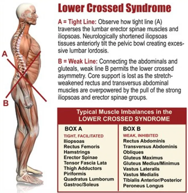
Lower Crossed Syndrome is a pattern of muscle imbalance involving the lumbar spine, pelvis, and hip. According to Das et al. (2017), this syndrome involves excessive tension in the hip flexors and lumbar extensors, along with weakness in the abdominal muscles and hip extensors. This pattern of movement primarily affects the L4-L5 and L5-S1 vertebral segments, the sacroiliac joint, and the hip joint.

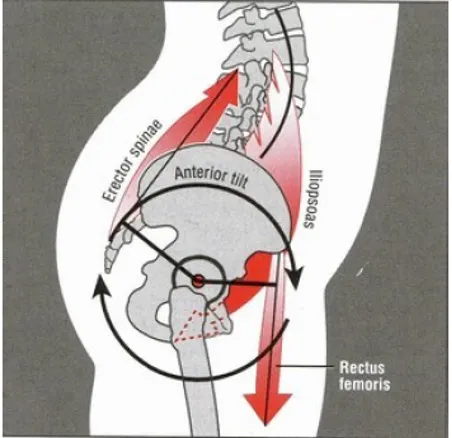
The change in muscle activation of the lumbopelvic-hip complex will lead to postural disorders that may increase the risk of injury and low back pain. The main change is an increase in anterior pelvic tilt, which causes hip flexion and increased lumbar lordosis, resulting in an excessive curve in the lower back (Kritz & Cronin, 2008). Other postural changes that may occur include lateral alterations of the pelvis, lateral rotation of the leg, or hyperextension of the knees.

SoccerDoc Blog EU

Image 1. Anterior pelvic tilt (Neumann, 2010)

More common than we think in football players

SoccerDoc Blog EU

Image 2. Rating scale: 5, ideal posture; 4, minor deviation; 3, significant deviation; 2, marked deviation; 1, severe deviation. (Kritz & Cronin, 2008)

Image 2 presents a study from 2008 by Kritz & Cronin, which demonstrated a tendency towards increased lumbar lordosis in athletes engaged in high-intensity running sports, such as football.
The study, involving 98 athletes, found that 33% of them exhibited marked or severe lumbar deviation during a static posture analysis. Authors like Das et al. (2017) have linked this type of postural imbalance to low back pain, while others, such as Mendiguchia et al. (2024),
have associated an increase in anterior pelvic tilt with greater elongation and strain in the hamstring muscle complex, leading to a higher risk of injury.

Football involves multiple explosive hip flexion movements, such as shooting the ball or passing, as well as short-duration, high-intensity actions like accelerations, decelerations, jumps, or changes of direction. These movements primarily stimulate the hip flexor and knee extensor muscles, leading to increased tension and overactivation of these muscle groups.

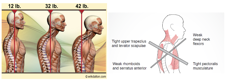
When one muscle contracts, the opposite or antagonist muscle relaxes. This is known as reciprocal inhibition. Excessively tight hip flexors will cause reciprocal inhibition of the gluteus maximus, the primary hip extensor (Buckthorpe, et al., 2019). This muscle activation pattern may cause excessive anterior pelvic tilt and, consequently, increase lumbar lordosis. In turn, the increase in lumbar lordosis will cause shortening and stiffness of the lower back muscles and reciprocal inhibition of the abdominal muscles (Image 3). Moreover, when the hip flexors become overactive, they start to take over functions that the abdominal muscles should perform. Again, this weakens the abdominal muscles and leads to the weakening and lengthening of the gluteus maximus due to their agonist-antagonist relationship (Das et al.,2017). Weakness in the gluteus maximus can lead to compensatory actions by secondary hip extensor muscles, such as the hamstrings and hip adductors, clinically known as “synergistic dominance” (Buckthorpe et al., 2019).

SoccerDoc Blog EU

Image 3. Lower crossed syndrome (Source: www.erikdalton.com)

How does this syndrome impact maximal speed running?

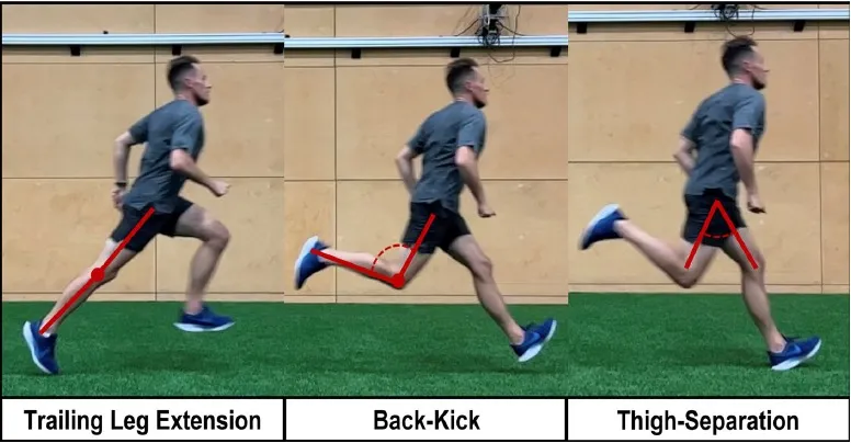
During maximum-speed running, athletes who present this motor pattern may increase hip and rear leg extension to compensate for the lower activation of the gluteus maximus (Image 4, left). Increasing hip extension during the final stance phase allows the athlete to apply force over a longer time, resulting in a greater impulse (Kritz & Cronin, 2008). Although this may seem positive, it’s actually an ineffective force production strategy used to compensate for the lack of force during the first half of the stance phase, where the peak of ground reaction forces should occur (Bramah et al., 2023). Excessive hip extension lengthens the stance phase and reduces the flight time and the time available to reposition the lower limb for the next foot contact, increasing the speed of the leg change and, consequently, the strain produced on the hamstrings (Bramah et al., 2023). Hip hyperextension may also cause excessive anterior pelvic tilt, which increases stress on the proximal hamstring region during high-intensity running (Mendiguchia et al., 2021).

SoccerDoc Blog EU

Image 4. Trailing leg extension (Bramah, et al., 2023)

Several authors, such as Schuermans et al. (2017), have shown how the increased activity of the hip flexors during sprinting increases the strain on the biceps femoris of the contralateral leg. This author also states that the activity of the abdominopelvic muscles, especially the gluteus maximus and abdominal obliques, reduces strain in the proximal region of this muscle. A proper balance of the lumbopelvic muscles, especially the relationship between iliopsoas-erector spinae and abdominal-gluteal muscles, is key to reducing strain on the biceps femoris (Mendiguchia et al., 2021).

This relationship once again highlights the importance of lumbopelvic control during maximum-intensity running. The increase in anterior pelvic tilt will cause a significant elongation of the hamstring muscles (Mendiguchia et al., 2024), which during high-intensity running increases the risk of suffering hamstring injuries (Mendiguchia et al., 2021).

Now that we understand the implications of this inefficient movement pattern, my next question is: How can we assess it and improve it?
We will address these questions in upcoming posts.

See you soon!

References

Bramah, C., Mendiguchia, J., Dos’Santos, T. & Morin, J., 2023. Exploring the role of sprint biomechanics in hamstring strain injuries: A current opinion on existing concepts and evidence. Sports Medicine, Volumen 54, pp. 783-793.

Buckthorpe, M., Stride, M. & Della Villa, F., 2019. Assessing and treating gluteus maximus weakness – A clinical commentary. The International Journal of Sports Physical Therapy, pp. 655-670.

Das, S. y otros, 2017. Prevalence of lower crossed syndrome in young adults: A cross sectional study. International Journal of Advanced Research, pp. 2217-2228.

Kritz, M. & Cronin, J., 2008. Static posture assessment screen of athletes: Benefits and considerations. Strength and Conditioning Journal, 30(5), pp. 18-27.

Mendiguchia, J. y otros, 2024. Anterior pelvic tilt increases hamstring strain and is a key factor to target for injury prevention and rehabilitation. Knee surgery, sports traumatology, arthroscopy , pp. 1-10.

Mendiguchia, J. y otros, 2021. Can we modify maximal speed running posture? Implications for performance and hamstring injury management. International Journal of Sports Physiology and Performance.

Neumann, D., 2010. Kinesiology of the musculoskeletal system. 2nd ed. Missouri: Mosby.

Improving Ankle Dorsiflexion: How to Do It?

VOETBAL MEDISCH SYMPOSIUM 2020

DE BEHANDELING VAN VOETBALBLESSURES 

PRAKTISCHE WETENSCHAP

OP DE KNVB CAMPUS IN ZEIST VINDT KOMEND JAAR OPNIEUW HET VOETBALMEDISCH SYMPOSIUM PLAATS.

HET SYMPOSIUM IS DÉ PLEK OM COLLEGA’S BINNEN HET VOETBALMEDISCHE DOMEIN TE ONTMOETEN OF KENNIS OP TE DOEN VAN GERENOMMEERDE EXPERTS. EN DIE NIEUWSTE INNOVATIES TE ZIEN OP HET GEBIED VAN VOETBALMEDISCHE EN FYSIEKE PRESTATIES.

NA VORIG JAAR DE DIAGNOSTIEK VAN VOETBALBLESSURES BELICHT TE HEBBEN, ROLT DE BAL DIT JAAR VERDER NAAR DE BEHANDELING VAN VOETBALBLESSURES. HET INHOUDELIJKE PROGRAMMA BIEDT OPNIEUW SPREKERS DIE ZICH ONDERSCHEIDEN IN ZOWEL DE DAGELIJKSE ZORG VOOR DE VOETBALLERS ALS OP WETENSCHAPPELIJK GEBIED.

Ja, ik wil mij aanmelden!

VOETBAL MEDISCHE WORKSHOP 2020

(VELD)REVALIDATIE NA EEN VOETBALBLESSURE

OP 4 MAART ZAL ER WEDEROM EEN WORKSHOP PLAATS VINDEN BIJ HET KNVB VOETBAL MEDISCH CENTRUM. 
OOK DIT JAAR BELOOFD HET EEN OCHTENDVULLEND PROGRAMMA TE ZIJN WAAR VOORNAMELIJK (SPORT)FYSIOTHERAPEUTEN HUN KENNIS MEE KUNNEN UITBREIDEN.

TIJDENS DE WORKSHOP ZAL MATT TABERNER ZIJN KENNIS EN EXPERTICE MET DE DEELNEMERS GAAN DELEN. MATT TABERNER IS EEN ERVAREN CLINICUS DIE AL JAREN EINDVERANTWOORDELIJK IS VOOR DE REVALIDATIE VAN TOPVOETBALLERS IN DE PREMIER LEAGUE. ZIJN FOCUS LIGT VOORNAMELIJK OP FYSIEKE ONTWIKKELING EN PRESTATIES. TEVENS IS HIJ DE ONTWIKKELAAR VAN HET ‘CONTROL-CHAOS CONTINUUM’.

DIT FRAMEWORK, WELKE VIJF FASES BESCHRIJFT HOE DE VELDREVALIDATIE NA EEN VOETBALBLESSURE OPGEBOUWD KAN WORDEN, STAAT CENTRAAL BINNEN DE WORKSHOP. DE THEORETISCHE ACHTERGROND,
DE TOEPASSING EN HET PRAKTISCHE ASPECT ZULLEN ALLEN AAN BOD KOMEN TIJDENS DE WORKSHOP.

JA, IK WIL MIJ AANMELDEN!

reviews

These guys are the best when it comes to style and have great attention to detail. 

Issac Newton
MODEL, FRANCE

These guys are the best when it comes to style and have great attention to detail. 

Issac Newton
MODEL, FRANCE

These guys are the best when it comes to style and have great attention to detail. 

Issac Newton
MODEL, FRANCE

Nick van der Horst

Meet the soccerdoc

Nick van der Horst behaalde zijn diploma fysiotherapie in 2007 aan de Hogeschool Utrecht. Hij werkte 10 jaar lang als sportfysiotherapeut/echografist/docent bij het Academie Instituut te Utrecht. Daarna heeft hij de overstap gemaakt naar waar zijn hart ligt, het professionele voetbal. Hij heeft twee jaar als sportfysiotherapeut en hoofd van de medische staf bij Go Ahead Eagles in Deventer gewerkt. Momenteel is is Nick werkzaam bij de KNVB. Zijn onderzoeks-activiteiten zijn gefocust op de voetbal-medische zorg. In 2017 behaalde hij zijn doctoraal na het verdedigen van zijn proefschrift ‘Prevention of hamstring injuries in male soccer’.

Blogger: Raúl Gómez
Blogger: Raúl Gómez

There are hundreds of variations of ankle mobility exercises on the internet, but while mobility training is an essential part of any training program, improving the range of motion (ROM) of a chronically restricted joint can be much more complex than doing a couple of stretches after training.

In the previous two articles, we discussed how limited ankle mobility can affect lower limb movement mechanics and explored various ankle dorsiflexion assessment methods. Now, it is time to design a training program for soccer players with ankle mobility restrictions.

In the book “Assessment and Treatment of Muscle Imbalance. The Janda Approach”, we find a very interesting classification of the musculature that tends to overactivation and the one that tends to weakness or inhibition. In the case of the ankle, the authors of this book state that the triceps surae muscles, especially the soleus, are prone to overactivation and stiffness, while other anterior lower leg muscles, such as the tibialis anterior, are prone to muscle weakness and inhibition. Authors such as Utku et al. (2018) have shown in their research the difference in the activation and inhibition patterns of the muscles of the triceps surae and the tibialis anterior. Their research showed stronger inhibition of the tibialis anterior compared to the triceps surae, which could be associated with specific neural control strategies of posture (Utku et al., 2018).

To address the muscular imbalance in the ankle joint, it is important to reduce the excessive tension and shortening of the posterior muscles while also activating and strengthening the anterior muscles. In a study by Lee et al. (2021), the effects of stretching the triceps surae alone were compared with the effects of combining the triceps surae stretching with tibialis anterior strengthening exercises (Image 1). The results showed that the group that performed the combination of triceps surae stretching and tibialis anterior strengthening increased ankle dorsiflexion ROM more than the group that performed stretching alone. After the exercises, the activity of the tibialis anterior muscle increased. The authors suggest that this increase could lead to reciprocal inhibition of the triceps surae musculature, thus increasing the ROM of the ankle. It is important to note that this study only looked at the immediate effects of the intervention, and there is very little research on the long-term effects of this type of intervention. Therefore, we do not know the chronic effects of applying this type of exercise.
Image1 SoccerDoc Blog EU Ankle

Image 1. Tibialis anterior resistance exercise

Static stretching has been proven to effectively improve flexibility and performance and reduce delayed-onset muscle soreness in the long term. However, it may have negative acute effects on muscle strength, potentially impairing sports performance (Grieve et al., 2022). On the other hand, alternative techniques like myofascial release have been shown to enhance the ROM without negatively affecting performance.

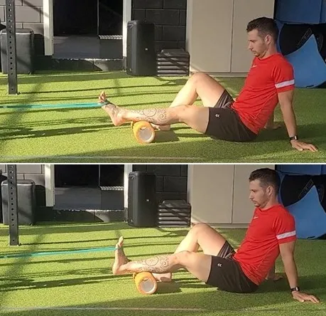
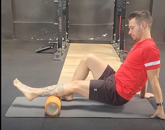
The use of the Foam Roller (FR) (Image 2) has gained much popularity in recent years, although many people are still unaware of its effects and how to use it properly. The review by Grieve et al. (2022) showed that the use of FR is effective in improving short-term ankle dorsiflexion ROM without detrimental effects on performance, making it a viable alternative to static stretching during warm-up. This improvement may be due to increased muscle temperature and pressure after using the FR. These changes would cause alterations in the density and fluidity of the muscle tissue (Aune et al., 2019). Other theories have also been suggested, such as the activation of the parasympathetic system after pressure is applied to the muscle. This activation would influence neural activity and reduce muscle tone (Aune et al., 2019).
Image 2 SoccerDoc Blog EU Ankle

Image 2. Self-myofascial release with Foam Roller

More research is needed regarding the long-term effects of FR since the investigations’ results are very different due to the significant difference between protocols. In the review previously mentioned, most studies use the FR for 1.5 to 3 minutes, applying as much pressure as possible. The tempo of FR varies significantly across studies, making it very difficult to draw conclusions about it.

Another alternative to static stretching is dynamic stretching, which would be done in the same positions as static stretching but moving through the full ROM at a set pace.

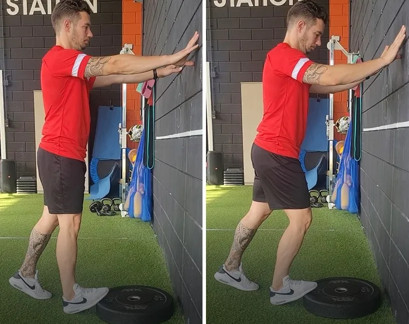
The inclusion of this type of stretching of the lower limb muscles, including exercises for gastrocnemius and soleus (Image 3), has shown not only short-term improvements in ankle mobility but also improvements in performance compared to a protocol that did not include dynamic soleus stretches (Huang et al., 2022).

Image 3 SoccerDoc Blog EU Ankle

Image 3. Gastrocnemius (left) and soleus stretching (right)

The results of these investigations suggest that combining FR with dynamic stretching may be a viable way to acutely improve ankle mobility and sports performance (Little & Williams, 2006).

This approach may be used during warm-ups prior to high-intensity sessions or competitions, although further research is necessary to determine the best way to apply both interventions.

Ankle dorsiflexion can also be limited by the restriction of posterior talus gliding, which is an essential movement for proper ankle joint motion (Jeon et al., 2015). To address this limitation in intra-articular mobility, mobilization techniques involving manual traction or straps have been suggested to assist with the posterior gliding of the talus during ankle dorsiflexion.

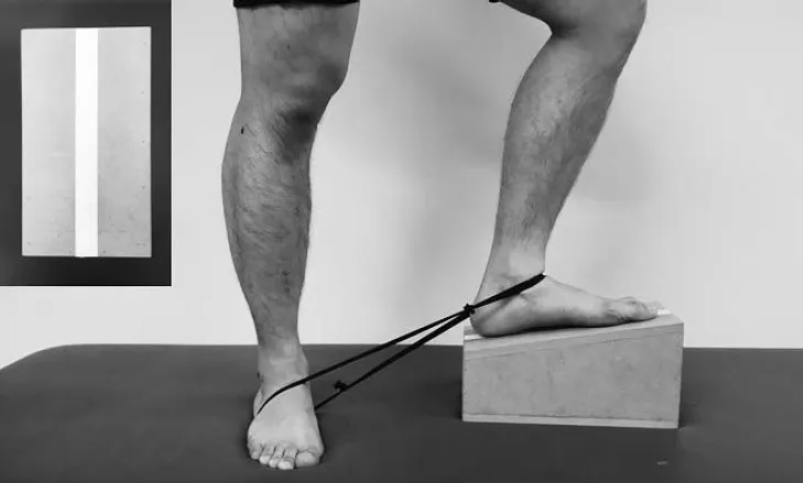
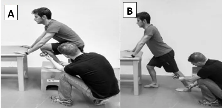
Jeon et al. (2015) compared the effect on ankle mobility of performing static triceps surae stretching alone with performing the same stretches using a strap that assisted posterior talus glide. In this technique, the strap was placed around the anterior aspect of the talus of the front foot, which was placed on a 10º inclined board. The other part of the strap was placed around the rear foot, which was on the ground, applying force on the front foot in a posteroinferior direction (Image 4).

Image 4 SoccerDoc Blog EU

Image 4. Ankle self-stretching using a strap (Jeon et al., 2015)

From this position, the patient was asked to move the knee forward without pain or discomfort until stretching of the soleus of the front leg was felt.
This final position was held for 20 seconds. 15 repetitions were performed, with a 10-second rest between stretches. This intervention was performed 5 times a week for 3 weeks in both groups. The group that performed stretching with the assistance of a strap showed better results compared to those who only did stretching in terms of active and passive ankle dorsiflexion and flexion angle during a closed kinetic chain test.

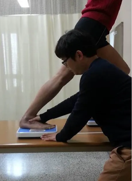
Similarly, Kang et al. (2015) examined the effects of combining static stretching with talocrural mobilization. In this case, the therapist assisted the posterior gliding of the talus with his hand (see Image 5). The stretches were performed in 10 repetitions of 30 seconds with 30 seconds of rest between repetitions.

This study also found superior effects of this type of mobilization compared to static stretching alone regarding increased ankle dorsiflexion, time to heel-off during gait, and posterior gliding of the talus.

Image 5 SoccerDoc Blog EU

Image 5. Manual talocrural mobilization (Kang et al., 2015)

Although eccentric exercise has been shown to increase muscle fascicle length effectively (Harris-Love et al., 2021), there is controversy surrounding its impact on improving ankle mobility. While authors such as Aune et al. (2019) have shown improvements following a training plan that included eccentric exercises, others such as Lagas et al. (2021) did not find any benefit; in fact, they observed a decrease in the flexibility of the triceps surae musculature.

This may be due to the difference between protocols. Lagas et al. (2021) performed the exercises 3 times per week following a progression that began with a very low training volume, 2 sets of 4 repetitions. The volume increased by about 20% each week until finishing the last week doing 3 sets of 15 repetitions. In the other study, Aune et al. (2019) performed the exercises daily for 4 weeks, starting directly with 3 sets of 15 and maintaining the same training volume throughout the intervention. Although eccentric training has been shown to be effective in improving muscle flexibility, more research is needed to determine the optimal training dose to improve ankle dorsiflexion ROM.

Certain medical conditions can limit joint ROM and need special clinical evaluation, but these cases are beyond the scope of this article.

In summary, a training plan aimed at improving ankle dorsiflexion ROM should include the following:
  • Myofascial release of the triceps surae
  • Talocrural mobilization with assistance to the posterior gliding of the talus
  • Stretching of the triceps surae (Dynamic stretching is recommended if performed before competition or training with high-intensity actions)
  • Specific strengthening of the dorsiflexor muscles
  • Eccentric exercises

Although isolated exercises can be very useful in improving certain risk factors for soccer injuries, we cannot forget that human movement is much more complex than the movement of a single joint. After correcting movement and strength deficits, sport-specific activities should be trained, including multi-articular exercises, plyometrics, and high-intensity actions such as sprinting, jumping, and changes of direction.

Injury prevention is a very complex process that requires daily effort. Achieving adequate mobility of all joints is critical to improving performance and minimizing the risk of severe injuries. As shown, the ankle joint plays a vital role in this process.

References

Aune, A. y otros, 2019. Acute and chronic effects of foam rolling vs eccentric exercise on ROM and force output of the plantar flexors. Journal of Sports Sciences, pp. 138-145.

Grieve, R. y otros, (2022). The effects of foam rolling on ankle dorsiflexion range of motion in healthy adults: A systematic literature review. Journal of Bodywork & Movement Therapies, pp. 53–59.

Harris-Love, M., Gollie, J. & Keogh, J. (2021). Eccentric Exercise: Adaptations and Applications for Health and Performance. Journal of functional morphology and kinesiology, 6(96).

Huang, S. y otros, 2022. Acute Effects of Soleus Stretching on Ankle Flexibility, Dynamic Balance and Speed Performances in Soccer Players. Biology, 11(374).

Jeon, I.-C.y otros, 2015. Ankle-Dorsiflexion Range of Motion After Ankle Self-Stretching Using a Strap. Journal of Athletic Training, 50(12), p. 1226–1232.

Kang, M.-H.y otros, 2015. Immediate combined effect of gastrocnemius stretching and sustained talocrural joint mobilisation in individuals with limited ankle dorsiflexion: A randomised controlled trial. Manual therapy, Volumen 20, pp. 827-834.

Lagas, I. F. y otros, 2021. Effects of eccentric exercises on improving ankle dorsiflexion in soccer players. BMC Musculoskeletal Disorders, 22(485).

Lee, J., Cynn, H., Shin, A. & Kim, B. (2021). Combined Effects of Gastrocnemius Stretch and Tibialis Anterior Resistance Exercise in Subjects with Limited Ankle Dorsiflexion. Physical Therapy Rehabilitation Science, 10(1), 10–15.

Little, T. & Williams, A., 2006. Effects of differential stretching protocols during warm-ups on high-speed motor capacities in professional soccer players. Journal of Strength and Conditioning Research, 20(1), pp. 203-207.

Page, P., Frank, C. & Lardner, R., (2012). Assessment and Treatment of Muscle Imbalance: The Janda Approach. Windsor, Ontario, Canada: Human Kinetics.

Utku, Y., Negro, F., Diedrichs, R. & Farina, D., 2018. Reciprocal inhibition between motor neurons of the tibialis anterior and triceps surae in humans. Journal of Neurophysiology, pp. 1699-1706.

Student blog: ACL rehabilitation in elite football

VOETBAL MEDISCH SYMPOSIUM 2020

DE BEHANDELING VAN VOETBALBLESSURES 

PRAKTISCHE WETENSCHAP

OP DE KNVB CAMPUS IN ZEIST VINDT KOMEND JAAR OPNIEUW HET VOETBALMEDISCH SYMPOSIUM PLAATS.

HET SYMPOSIUM IS DÉ PLEK OM COLLEGA’S BINNEN HET VOETBALMEDISCHE DOMEIN TE ONTMOETEN OF KENNIS OP TE DOEN VAN GERENOMMEERDE EXPERTS. EN DIE NIEUWSTE INNOVATIES TE ZIEN OP HET GEBIED VAN VOETBALMEDISCHE EN FYSIEKE PRESTATIES.

NA VORIG JAAR DE DIAGNOSTIEK VAN VOETBALBLESSURES BELICHT TE HEBBEN, ROLT DE BAL DIT JAAR VERDER NAAR DE BEHANDELING VAN VOETBALBLESSURES. HET INHOUDELIJKE PROGRAMMA BIEDT OPNIEUW SPREKERS DIE ZICH ONDERSCHEIDEN IN ZOWEL DE DAGELIJKSE ZORG VOOR DE VOETBALLERS ALS OP WETENSCHAPPELIJK GEBIED.

Ja, ik wil mij aanmelden!

VOETBAL MEDISCHE WORKSHOP 2020

(VELD)REVALIDATIE NA EEN VOETBALBLESSURE

OP 4 MAART ZAL ER WEDEROM EEN WORKSHOP PLAATS VINDEN BIJ HET KNVB VOETBAL MEDISCH CENTRUM. 
OOK DIT JAAR BELOOFD HET EEN OCHTENDVULLEND PROGRAMMA TE ZIJN WAAR VOORNAMELIJK (SPORT)FYSIOTHERAPEUTEN HUN KENNIS MEE KUNNEN UITBREIDEN.

TIJDENS DE WORKSHOP ZAL MATT TABERNER ZIJN KENNIS EN EXPERTICE MET DE DEELNEMERS GAAN DELEN. MATT TABERNER IS EEN ERVAREN CLINICUS DIE AL JAREN EINDVERANTWOORDELIJK IS VOOR DE REVALIDATIE VAN TOPVOETBALLERS IN DE PREMIER LEAGUE. ZIJN FOCUS LIGT VOORNAMELIJK OP FYSIEKE ONTWIKKELING EN PRESTATIES. TEVENS IS HIJ DE ONTWIKKELAAR VAN HET ‘CONTROL-CHAOS CONTINUUM’.

DIT FRAMEWORK, WELKE VIJF FASES BESCHRIJFT HOE DE VELDREVALIDATIE NA EEN VOETBALBLESSURE OPGEBOUWD KAN WORDEN, STAAT CENTRAAL BINNEN DE WORKSHOP. DE THEORETISCHE ACHTERGROND,
DE TOEPASSING EN HET PRAKTISCHE ASPECT ZULLEN ALLEN AAN BOD KOMEN TIJDENS DE WORKSHOP.

JA, IK WIL MIJ AANMELDEN!

reviews

These guys are the best when it comes to style and have great attention to detail. 

Issac Newton
MODEL, FRANCE

These guys are the best when it comes to style and have great attention to detail. 

Issac Newton
MODEL, FRANCE

These guys are the best when it comes to style and have great attention to detail. 

Issac Newton
MODEL, FRANCE

Nick van der Horst

Meet the soccerdoc

Nick van der Horst behaalde zijn diploma fysiotherapie in 2007 aan de Hogeschool Utrecht. Hij werkte 10 jaar lang als sportfysiotherapeut/echografist/docent bij het Academie Instituut te Utrecht. Daarna heeft hij de overstap gemaakt naar waar zijn hart ligt, het professionele voetbal. Hij heeft twee jaar als sportfysiotherapeut en hoofd van de medische staf bij Go Ahead Eagles in Deventer gewerkt. Momenteel is is Nick werkzaam bij de KNVB. Zijn onderzoeks-activiteiten zijn gefocust op de voetbal-medische zorg. In 2017 behaalde hij zijn doctoraal na het verdedigen van zijn proefschrift ‘Prevention of hamstring injuries in male soccer’.

Blogger: Cathal Corr

SoccerDoc stimulates knowledge development and personal development of anyone working within the field of Football Medicine. In addition to our bloggers who provide solid evidence-based coverage of certain topics,
we provide a platform for students to submit their work and share it with the SoccerDoc-community.
Feel free to share any knowledge, experiences and or comments below the blog to learn from each other.

Introduction

Anterior cruciate ligament injuries rank among the most prevalent and devastating injuries, affecting both amateur and elite athletes (van Melick, 2016). Despite the advancements in research and clinical practice, players playing at a professional level have a recurrence rate of 17.8% (Wright et al., 2007). Despite notable strides in research and clinical practices, professional players face a concerning recurrence rate of 17.8% (Wright et al., 2007).
Moreover, rerupture rates post-reconstruction range from 3-22% on the reconstructed ligament and 3-24% on the contralateral side, further complicating the rehabilitation process. The risk of a subsequent second ACL tear is not the only barrier to a successful return to sport, the return to preinjury level participation post ACL surgery is reported to be as low as 65%, underlining the multifaceted challenges athletes encounter in their journey back to peak performance (Ardern et al., 2015).

Against this backdrop, this blog endeavours to explore strategies for optimising ACL rehabilitation within the context of a multidisciplinary team (MDT) working within an elite football team setting. By delving into the workings of the MDT, we aim to elucidate how proactive organisational structures, effective rehabilitation planning, and open communication channels can bolster the success of rehabilitation efforts. Through these avenues, we seek to empower medical teams to navigate the complexities of ACL rehabilitation ultimately fostering the return-to-performance of the athletes with confidence and resilience.

Operational Structure

In elite football, a typical multidisciplinary team comprises five distinct departments, each playing a vital role in the management and preparation of players: coaches, medical team, performance team, data science team, and analysis team (Transfermarkt, 2023).

Each department brings specialised expertise and responsibilities to the table, collectively contributing to the optimisation of player performance and well-being.

Image1

Image 1. Multidisciplinary Team

Let’s briefly outline the roles and responsibilities associated with the two most prominent departments involved with the rehabilitation of a player with an ACL tear:

The Medical Department

The medical department consists of a sports medicine physician (club doctor) and typically 2-3 physiotherapists.

The Sports Medicine Physician
The sports medicine physician is typically the case manager and will oversee the rehabilitation period (Villa et al., 2019).

The Physiotherapist
The physiotherapists in the MDT will be responsible for the majority of the ACL rehabilitation. The regaining of range of motion, strength, power, hypertrophy, balance, and coordination are well-established and researched goals for the athlete to achieve under the guidance of the MDT; specifically the physiotherapist (Taberner et al., 2020).

The Performance Department

The performance department has a vital role to play in the reconditioning of the athlete back to the pre-injury level.

The strength and conditioning coach(s)
Here, the strength and conditioning coaches will reintroduce upper-body training and more dynamic and full-body movements once the initial protective phase has been completed (add more detail) at approximately 2-3 months post-surgery. The reconditioning of the body is crucial to regain the physical profile to compete at the highest level but also to build resilience to mitigate any further injuries (Bizzini et al., 2012).

The sports psychologist
However, the reconditioning of the mind is equally important and can be less straightforward (Ardern, 2015). Often overlooked, the psychological aspect of an ACL injury in an elite footballer cannot be underestimated. Mental training techniques to manage pain and reduce anxiety associated with injury are essential (Clement &Arvinen-Barrow, 2013). The sport psychologist provides screening and support for the player, with early identification of psychological barriers being crucial. Specific questionnaires to assess athletes’ fear, confidence, and perceptions of risk can help tailor interventions to their needs. This proactive approach can prevent psychological issues from hindering physical recovery (Ardern et al., 2013). Setting specific, measurable, achievable, relevant, and time-bound (SMART) goals helps athletes build confidence and stay motivated during their recovery (Ardern et al., 2022). The psychologist also encourages autonomy and control by advising the player to take an active role in their rehabilitation alongside the medical staff. Positive mental imagery, such as visualisation techniques, helps athletes imagine a successful return to the football pitch, reducing anxiety and fostering a positive mindset toward recovery and performance (Ardern et al., 2013). ACL rehabilitation is not a straightforward process, and setbacks are inevitable. The sport psychologist helps athletes develop coping strategies, emphasising relaxation exercises, cognitive-behavioural strategies, and ensuring adequate social support (Ardern et al., 2022).

Furthermore, it is my opinion that both the medical department and performance department may help with easing uncertainty, doubt, and fear of re-injury from the start. The medical and performance staff’s plan should give the players objective and measurable targets to hit. Players are natural competitors, and a long-term ACL injury rips away a big part of their life that motivates them daily. Measurable and objective short-term goals and targets can help feed that competitive urge and motivate the player. In addition to this, speaking with another player who has gone through the same long-term injury can allow the sharing of experiences, challenges, and solutions that we as non-playing staff members can not relate to.

Image2

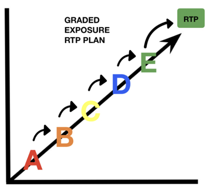
Image 2. Graded Exposure RTP Plan

Planning

A return to sport training plan should be completed within the first week of the ACL reconstruction. This plan should be coordinated and led by the ‘case manager’ (CM). The CM is typically the sports medicine physician within the organisation that leads the rehabilitation from start to finish. Their role is to give strategic and practical advice to the MDT on a daily basis and in key decision-making throughout the recovery process. When surgery is needed, the case manager liaises with the surgeon regarding the stages of tissue healing and the appropriate loading of the knee post-surgery (Villa et al., 2019). There are logistical factors such as travel requirements, budget, resources, MDT meeting schedule, and testing schedule. The recovery timeline should be forecast by taking into consideration the datasets of previous players who underwent ACL surgery. However, some covariates that need to be taken into consideration are previous knee injuries, the time of the season, and concurrent injuries such as a chondral injury, a meniscal tear or multi-ligament injuries (Jordan et al., 2015).

From my personal experience as a practitioner, a plan with clear milestones and progression/regression criteria enables the athlete to have a bird’s eye view of the rehabilitation plan and can help with any doubts or fears they may have. ACL rehabilitation is a long journey and there may be setbacks along the way.

Communication

Acute stage
Acute injuries such as an ACL tear challenge the resilience and organisation of the MDT and emphasise the importance of teamwork and communication in such situations. On match days, at least one team doctor and at least one senior physiotherapist will make up the medical team on the sideline (The Football Association, 2023). In the event of an ACL tear, the medical team needs to be well-equipped to handle this scenario by coordinating their efforts effectively and following a clear and pre-planned protocol. Below I have illustrated the process of coordinating an ACL tear in training or on a match day.

Acute Injury protocol:

Image3

Image 3. Acute ACL Injury Protocol

Following the receipt of the imaging report, the CM follows a carefully controlled protocol. This allows discussion and communication of the injury, initially with one decision-maker, then with the Head Physio, a specialist if relevant, with the player, with the manager, director of football, medical team, logistics team and finally the board. Crucially, any media announcements should only be made after all the above individuals and teams have been informed and discussed the injury (Ahmad, n.d).

Use of Whatsapp

The day-to-day life of a footballer when not injured is carefully laid out and structured around team training, gym, tactical sessions and recovery. When our athlete is undergoing ACL rehabilitation, we should try to minimise the change of routine and help the athlete adjust to their new routine. The use of WhatsApp groups for rehabilitation can allow members of the medical team to quickly and effectively transfer information to athletes about training programmes, at-home rehabilitation and logistics around when and where to be day to day. Additionally, WhatsApp allows files, photos, and videos to be shared and stored for future reference. This can be of great benefit in the initial weeks post-surgery, as many players will travel abroad to undergo their surgery with their desired surgeon. WhatsApp messaging allows the player to relay information regarding their surgery, the status of their knee such as swelling, pain and range of motion and their overall well-being to the staff daily. It is important to note that this form of communication does not replace standard check-ins via video call or phone, but instead enhances the communication in between check-ins. Due to the understandable anxiety players feel around more sensitive information such as x-ray results and MRI scan results, this sharing of information should ideally be done in person or on the phone. This is not the purpose of the WhatsApp group. Continuous and effective communication is warranted within the caregivers’ team. Having a communication model (e.g., digital clinical records) and periodic (daily if necessary) meetings are the milestones of a well-organised service. Formal and informal communication is key to enhancing the relationship between the different players. The more you communicate within the team, the more you can maximise the patient or athlete’s perception of the recovery path and minimise the risk of misleading information to the patient (Ahmed et al., 2020). The use of WhatsApp groups does however bring the privacy of the patient and the ethical dilemma of sharing private medical information. Depending on the country, the laws around this topic vary, thus ensuring you are in line with your governing body and the specific privacy laws in your jurisdiction.

Conclusion
In conclusion, optimising ACL rehabilitation in professional football requires a comprehensive approach integrating structured multidisciplinary teamwork, meticulous planning, and open communication channels. By establishing clear operational structures within the multidisciplinary team, defining staff roles and responsibilities, and implementing effective planning strategies, such as coordinating return-to-sport training plans early on, the rehabilitation process can be streamlined for optimal outcomes. Moreover, leveraging communication tools like WhatsApp enhances the flow of information between medical staff and athletes, fostering a supportive environment crucial for successful recovery. With these strategies in place, the goal of leaving no stone unturned in ACL rehabilitation within elite football becomes more achievable, ensuring athletes can return to the field with confidence and resilience.

References

Football club doctor – An evolving role – injury rehab network event. Sterosport, https://sterosport.co.uk/dr-imtiaz-ahmad-football-club-doctor-an-evolving-role/

Ahmed, O. H., Carmody, S., Walker, L., & Ahmad, I. (2020). The need for speed! 10 ways that WhatsApp and instant messaging can enhance communication (and clinical care) in sports and exercise medicine. British Journal of Sports Medicine, 54(19), 1128-1129. https://doi.org/10.1136/bjsports-2019-101707

Ardern, C. L. (2015). Anterior cruciate ligament reconstruction—not exactly a one-way ticket back to the pre-injury level. Sports Health: A Multidisciplinary Approach, 7(3), 224-230. https://doi.org/10.1177/1941738115578131

Ardern, C. L., Hooper, N., O’Halloran, P., Webster, K. E., & Kvist, J. (2022). A psychological support intervention to help injured athletes “get back in the game”: design and development study. JMIR Formative Research, 6(8), e28851. https://doi.org/10.2196/28851

Ardern, C. L., Taylor, N. F., Feller, J. A., & Webster, K. E. (2013). A systematic review of the psychological factors associated with returning to sport following injury. British journal of sports medicine, 47(17), 1120–1126. https://doi.org/10.1136/bjsports-2012-091203

Arsenal FC – Staff. (n.d). Transfermarkt. Retrieved February 5, 2024, from https://www.transfermarkt.com/fc-arsenal/mitarbeit/verein/11

Bizzini, M., Hancock, D., & Impellizzeri, F. M. (2012). Suggestions from the field for return to sports participation following anterior cruciate ligament reconstruction: soccer. Journal of Orthopaedic & Sports Physical Therapy, 42(4), 304-312. https://doi.org/10.2519/jospt.2012.4005

Brinlee, A. W., Dickenson, S., Hunter‐Giordano, A., & Snyder‐Mackler, L. (2021). Acl reconstruction rehabilitation: clinical data, biologic healing, and criterion-based milestones to inform a return-to-sport guideline. Sports Health: A Multidisciplinary Approach, 14(5), 770-779. https://doi.org/10.1177/19417381211056873

Christakou, Α. and Lavallee, D. (2009). Rehabilitation from sports injuries: from theory to practice. Perspectives in Public Health, 129(3), 120-126. https://doi.org/10.1177/1466424008094802

Clement, D., & Arvinen-Barrow, M. (2013). Sport medicine team influences in psychological rehabilitation: A multidisciplinary approach. In M. Arvinen-Barrow & N. Walker (Eds.), The psychology of sport injury and rehabilitation(pp. 156–170). Routledge/Taylor & Francis Group.

Football Association. (2023). Premier League Medical regulations. Appointment of medical personnel, 2.1. theFA.com. https://www.thefa.com › rules-of-the-association

Jordan, M. J. and Aagaard, P. (2017). Anterior cruciate ligament injury/reinjury in alpine ski racing: a narrative review. Open Access Journal of Sports Medicine, Volume 8, 71-83. https://doi.org/10.2147/oajsm.s106699

Taberner, M., Dyk, N. v., Allen, T., Jain, N., Richter, C., Drust, B., … & Cohen, D. D. (2020). Physical preparation and return to performance of an elite female football player following ACL reconstruction: a journey to the FIFA women’s World Cup. BMJ Open Sport & Exercise Medicine, 6(1), e000843. https://doi.org/10.1136/bmjsem-2020-000843

Van Melick, N., Van Cingel, R., Brooijmans, F., Neeter, C., Van Tienen, T. G., Hullegie, W., & Sanden, M. W. N. D. (2016). Evidence-based clinical practice update: practice guidelines for anterior cruciate ligament rehabilitation based on a systematic review and multidisciplinary consensus. British Journal of Sports Medicine, 50(24),1506–1515. https://doi.org/10.1136/bjsports-2015-095898

Villa, F., Villa, S., & Mendes, J. E. (2019). Multidisciplinary sports medicine team. The Sports Medicine Physician, 3-11. https://doi.org/10.1007/978-3-030-10433-7_1

Wright, R. W., Dunn, W. R., Amendola, A., Andrish, J. T., Bergfeld, J. A., Kaeding, C. C., … & Spindler, K. P. (2007). Risk of tearing the intact anterior cruciate ligament in the contralateral knee and rupturing the anterior cruciate ligament graft during the first 2 years after anterior cruciate ligament reconstruction. The American Journal of Sports Medicine, 35(7), 1131-1134. https://doi.org/10.1177/0363546507301318

Ankle Dorsiflexion Mobility Assessment

VOETBAL MEDISCH SYMPOSIUM 2020

DE BEHANDELING VAN VOETBALBLESSURES 

PRAKTISCHE WETENSCHAP

OP DE KNVB CAMPUS IN ZEIST VINDT KOMEND JAAR OPNIEUW HET VOETBALMEDISCH SYMPOSIUM PLAATS.

HET SYMPOSIUM IS DÉ PLEK OM COLLEGA’S BINNEN HET VOETBALMEDISCHE DOMEIN TE ONTMOETEN OF KENNIS OP TE DOEN VAN GERENOMMEERDE EXPERTS. EN DIE NIEUWSTE INNOVATIES TE ZIEN OP HET GEBIED VAN VOETBALMEDISCHE EN FYSIEKE PRESTATIES.

NA VORIG JAAR DE DIAGNOSTIEK VAN VOETBALBLESSURES BELICHT TE HEBBEN, ROLT DE BAL DIT JAAR VERDER NAAR DE BEHANDELING VAN VOETBALBLESSURES. HET INHOUDELIJKE PROGRAMMA BIEDT OPNIEUW SPREKERS DIE ZICH ONDERSCHEIDEN IN ZOWEL DE DAGELIJKSE ZORG VOOR DE VOETBALLERS ALS OP WETENSCHAPPELIJK GEBIED.

Ja, ik wil mij aanmelden!

VOETBAL MEDISCHE WORKSHOP 2020

(VELD)REVALIDATIE NA EEN VOETBALBLESSURE

OP 4 MAART ZAL ER WEDEROM EEN WORKSHOP PLAATS VINDEN BIJ HET KNVB VOETBAL MEDISCH CENTRUM. 
OOK DIT JAAR BELOOFD HET EEN OCHTENDVULLEND PROGRAMMA TE ZIJN WAAR VOORNAMELIJK (SPORT)FYSIOTHERAPEUTEN HUN KENNIS MEE KUNNEN UITBREIDEN.

TIJDENS DE WORKSHOP ZAL MATT TABERNER ZIJN KENNIS EN EXPERTICE MET DE DEELNEMERS GAAN DELEN. MATT TABERNER IS EEN ERVAREN CLINICUS DIE AL JAREN EINDVERANTWOORDELIJK IS VOOR DE REVALIDATIE VAN TOPVOETBALLERS IN DE PREMIER LEAGUE. ZIJN FOCUS LIGT VOORNAMELIJK OP FYSIEKE ONTWIKKELING EN PRESTATIES. TEVENS IS HIJ DE ONTWIKKELAAR VAN HET ‘CONTROL-CHAOS CONTINUUM’.

DIT FRAMEWORK, WELKE VIJF FASES BESCHRIJFT HOE DE VELDREVALIDATIE NA EEN VOETBALBLESSURE OPGEBOUWD KAN WORDEN, STAAT CENTRAAL BINNEN DE WORKSHOP. DE THEORETISCHE ACHTERGROND,
DE TOEPASSING EN HET PRAKTISCHE ASPECT ZULLEN ALLEN AAN BOD KOMEN TIJDENS DE WORKSHOP.

JA, IK WIL MIJ AANMELDEN!

reviews

These guys are the best when it comes to style and have great attention to detail. 

Issac Newton
MODEL, FRANCE

These guys are the best when it comes to style and have great attention to detail. 

Issac Newton
MODEL, FRANCE

These guys are the best when it comes to style and have great attention to detail. 

Issac Newton
MODEL, FRANCE

Nick van der Horst

Meet the soccerdoc

Nick van der Horst behaalde zijn diploma fysiotherapie in 2007 aan de Hogeschool Utrecht. Hij werkte 10 jaar lang als sportfysiotherapeut/echografist/docent bij het Academie Instituut te Utrecht. Daarna heeft hij de overstap gemaakt naar waar zijn hart ligt, het professionele voetbal. Hij heeft twee jaar als sportfysiotherapeut en hoofd van de medische staf bij Go Ahead Eagles in Deventer gewerkt. Momenteel is is Nick werkzaam bij de KNVB. Zijn onderzoeks-activiteiten zijn gefocust op de voetbal-medische zorg. In 2017 behaalde hij zijn doctoraal na het verdedigen van zijn proefschrift ‘Prevention of hamstring injuries in male soccer’.

Blogger: Raúl Gómez
Blogger: Raúl Gómez

The previous article demonstrated how restricted ankle dorsiflexion can impact landing mechanics by increasing ground reaction forces and causing medial knee displacement (Taylor et al., 2021). If you haven’t read it yet, we recommend that you do so in order to understand the importance of ensuring adequate ankle mobility in football players. Read here.

Football is a high-intensity sport with a high number of eccentric muscle actions that occur during changes of direction, jumps or decelerations. Continuous exposure to these types of movement may increase the tightness of the muscles and tendons and cause muscle damage, which will affect the neural properties of the myotendinous unit, reducing the total range of motion throughout the season (Moreno- Pérez et al., 2020), increasing the risk of injury (Mason-Mackay et al., 2017) and negatively influencing sports performance (Gonzalo-Skok et al., 2015).

Although there is general agreement that the lack of ankle dorsiflexion increases the risk of lower limb injuries (Mason-Mackay et al., 2017), there is a notable difference in the evaluations utilized in various scientific articles and in the cut-off values for each test. In addition, there is no consensus on the optimal amount of mobility of this joint and how (lack of) mobility is associated with injury risk.

The tests used to assess ankle mobility can be classified into three categories:

  • Open Kinetic Chain (OCC)
  • Closed Kinetic Chain (CCC)
  • Landing mechanics

Different methods have been used to measure the results of each test, such as a goniometer, inclinometer, or analysis of digital models. In this article, we will not discuss the measurement method.

Open Kinetic Chain (OKC)

This type of evaluation is the least functional to sports activity since all the actions where ankle mobility is important are performed in closed kinetic chains (landings, changes of direction, running, etc.). Although this type of test will not provide us with any information about the control and mobility that the football player has in specific actions, it can be very useful when evaluating mobility restrictions or pain on specific structures.

In these tests, ankle dorsiflexion is evaluated with the patient sitting or lying on the treatment table. The ankle is passively dorsiflexed until the limit of movement is reached. This test should be performed first with the knee extended to assess flexibility of the gastrocnemius muscle, and then with the knee flexed at 90º to assess flexibility of the soleus. (Stovitz & Coetzee, 2004). Stovitz & Coetzee (2004) recommend testing this musculature with the subtalar joint in a neutral position while lateral force is applied to the neck of the talus and the forefoot is pushed medially (Image 1).

In this way, the foot is locked, preventing the eversion of the calcaneus, which could give a false impression of greater flexibility. Ankle dorsiflexion can be limited not only by restrictions in muscle flexibility, but also by intra-articular factors such as tightness of the talocrural posterior capsule, positional faults of the fibula, or reduced posterior glide of the talus relative to the ankle mortise (Howe, 2020).

SoccerDoc Blogs

Image 1. OKC ankle mobility assessment. (Stovitz & Coetzee, 2004)

Several authors recommend that the range of motion in these tests should reach 20º of dorsal flexion (Brockett & Chapman, 2016) (Magee, 2014). Stiffler et al. (2014), compared range of motion values during this type of test in individuals who do not show medial knee displacement during an overhead squat compared to those who do (Image 2). Individuals with greater ankle dorsiflexion range of motion showed less medial knee displacement.

SoccerDoc Blog

Image 2. Range of motion in OKC tests in the study by Stiffler et al. (2014); MKD – Medial knee displacemen

Due to the significant variation among studies, it is challenging to propose cut-off values. It is essential to compare both legs and ensure the absence of unilateral deficits. There is not much reference data on this type of evaluation in soccer players.

Closed Kinetic Chain (CKC)

In comparison to OKC tests, CKC assessments provide more information about ankle joint mobility in positions similar to those that occur during sports activities. Movement deficits during this type of test have been related to movement alterations that increase the risk of injury, such as knee valgus, excessive hip adduction, or greater trunk inclination (Ivana Hanzlíková et al., 2022).

There are many variations of CKC assessment. One test that is frequently applied is the Weight Bearing Lunge Test (WBLT) or Wall Test (Image 3). In this test, the subject is placed in a lunge position facing a wall, with one leg in front of the other. From this position, the patient lunges forward until the knee touches the wall, keeping the heel on the floor. If there is contact with the wall, the foot is moved back until finding the farthest distance where the knee contacts the wall while the heel remains entirely in contact with the ground. When the limit of movement is reached, the therapist measures the distance from the wall to the tip of the big toe.

SoccerDoc Blog

Image 3. Weight Bearing Lunge Test or Wall test

There are variations of this test, such as the Leg Motion System Test (Moreno-Pérez et al., 2020) or the Half Kneeling Dorsiflexion Test (Gourlay et al., 2019), but they all consist of the same movement, only varying the measuring device or the position of the rear leg. 

Dill et al. (2014) suggested a cutoff value of 44.02º for this test, while other authors, such as Moreno-Pérez et al. (2020), did not specify cutoff values in their studies but indicated that differences of more than 2 cm between legs should be considered as asymmetries that increase the risk of injury. Once again, it is challenging to establish exact cutoff values for soccer players due to the wide range of results in different studies.

The Modified Weight-Bearing Lunge Test, also known as the Ankle Clearing Test (Image 4), is performed with the patient standing with one leg in front of the other. The heel of the front leg is in contact with the toes of the back leg. A dowel is given to the patient to help maintain balance. From this position, the patient moves the knee of the rear leg forward as far as possible, keeping the heel in contact with the ground. 

The therapist measures the position of the knee in relation to the medial malleolus of the ankle of the front leg: behind, in line, or beyond (Gourlay, et al., 2019). This test has been shown to be a valid tool in evaluating ankle dorsiflexion and has been compared with the values obtained in the WBLT. 

The modified WBLT score corresponds to 33.5 ± 2.0 degrees if the knee is behind the medial malleolus of the front leg, 38.6 ± 1.2 degrees if it is in line, and 43.0 ± 0.78 degrees if it is beyond (Gourlay, et al., 2019). Therefore, soccer players with adequate ankle mobility should be able to move the knee beyond the medial malleolus of the front leg.

SoccerDoc Blog

Image 4.
Modified Weight Bearing Lunge Test or Ankle clearing test

Image 5 shows two other ankle dorsiflexion tests. One with the knee in extension (Right, the gastrocnemius will limit the result of this test to a greater extent) and another with the knee in flexion (Left, the soleus will limit the result of this test to a greater extent).

SoccerDoc Blog

Image 5.
Tests variations for the measurement of ankle dorsiflexion. Knee in flexion (Front leg; Left); Knee Extension (Back Leg; Right)

López-Valenciano et al. (2019), in a study with professional soccer players, used cut-off values of <17º for the test with the knee in extension and &<34º for the test with the knee in flexion. Lower values in these tests would be considered movement restrictions, increasing the risk of lower limb injuries.

Landing Mechanics

Various types of landing mechanics assessment have been used to assess lower limb movement mechanics. To summarize, we propose three groups: Bilateral, unilateral, and jump after landing. Once again, due to the wide variety in methodology (such as jump height, measurement method, and instructions during landing), it is challenging to propose standard tests and cut-off values.

SoccerDoc Blog

Image 6. Unilateral and bilateral landing

One of the most used tests in research is the Landing Error Scoring System” (LESS) test. It is performed from a 30 cm high box, from which the subject jumps forward at a distance equivalent to 50% of the subject’s height. In this test, the landing is bilateral. The system utilizes a 17-item scoring method to identify athletes who are at risk of injury. It is used to assess the ankle and the entire lower limb. In a study conducted by Padua et al. (2015), an example is provided to demonstrate how this evaluation method can be applied to soccer players. You can find the details in the references.

Whitting et al. (2011), conducted a study based on single-leg drop landings from heights of 32 and 72 cm. Malloy et al. (2014) assessed landing mechanics by having participants perform a maximum vertical jump immediately after landing from a box jump, with the height of the box matching the maximum height reached in a previous vertical jump by each participant. In both studies, researchers observed altered movement patterns in participants with limited ankle mobility measured by static tests.

Conclusion
OKC evaluations can help us detect deficits in isolated structures in the early stages of rehabilitation. However, once we have corrected the deficits, we must progress to tests and exercises with more functionality (e.g. CKC tests) and ensure the player is ready for competition.
The CKC tests are a quick and effective way to assess ankle mobility and identify soccer players at higher risk of injury. This makes them a good option when planning injury prevention programs. However, there is debate about whether static assessments are useful in predicting injuries. Even if a football player has good ankle mobility, we don’t know if they use an appropriate landing strategy. There may also be other deficits that predispose the football player to a higher risk of injury, such as excessive knee valgus due to weakness of the gluteal muscles, lack of lumbopelvic control or poor ankle stiffness.
Evaluating the landing mechanics of soccer players who have suffered long-term injuries, such as to the anterior cruciate ligament, or who experience chronic pain, for example, in the Achilles tendon or patellofemoral area, can provide essential information before they finish their rehabilitation and return to train or match play.
In our upcoming blog, the last of this series, we will explore different methods to enhance ankle mobility. We will identify which structures limit the movement and which ones are weakened or inhibited and propose targeted mobilizations and exercises to improve ankle dorsiflexion.

References

Brockett, C., & Chapman, G. (2016). Biomechanics of the ankle. Orthopaedics and trauma, 30(3), 232-238.

Dill, K., Begalle, R., Frank, B., Zinder, S., & Padua, D. (2014). Altered Knee and Ankle Kinematics During Squatting in Those With Limited Weight-Bearing–Lunge Ankle Dorsiflexion Range of Motion. Journal of Athletic Training, 49(6), 723-732.

Gonzalo-Skok, O., Serna, J., Rhea, M. R., & Marin, P. J. (2015). Relationships between Functional Movement Tests and Performance Tests in Young Elite Male Basketball Players. International Journal of Sports Physical Therapy, 10(5), 628-638.

Gourlay, J., Bullock, G., Weaver, A., Matsel, K., Kiesel, K., & Plisky, P. (2019). The Reliability and Criterion Validity of a Novel Dorsiflexion Range of Motion Screen. Athletic Training and Sports Health Care, 12(1).

Howe, L. P. (2020). Restrictions in ankle dorsiflexion range of motion and its effect on landing mechanics. Thesis, Ede Hill University.

Ivana Hanzlíková, I., Richards, J., & Hébert Losier, K. (2022). The influence of ankle dorsiflexion range of motion on unanticipated cutting kinematics. Sport Sciences for Health.

López-Valenciano, A., Ayala, F., Vera-García, F., De Ste Croix, M., Hernández, S., Ruiz, I., Cejudo, A.,  Santonja, F. (2019). Comprehensive profile of hip, knee and ankle ranges of motion in professional football players. The Journal of Sports Medicine and Physical Fitness, 59(1), 102-109.

Magee, D. (2014). Orthopedic Physical Assessment (Sixth ed.). St Louis, Missouri: Elsevier.

Malloy, P., Meinerz, C., Geiser, C., & Kipp, K. (2015). The Association of Dorsiflexion Flexibility on Landing Mechanics during a Drop Vertical Jump. Knee Surgery, Sports Traumatology, Arthroscopy, 23(12).

Mason-Mackay, A., Whatman, C., & Reid, D. (2017). The effect of reduced ankle dorsiflexion on lower extremity mechanics during landing: A systematic review. Journal of Science and Medicine in Sport, 20, 451–458.

Moreno-Pérez, V., Soler, A., Ansa, A., López-Samanes, Á., Madruga-Parera, M., Beato, M., & Romero-Rodríguez, D. (2020). Acute and chronic effects of competition on ankle dorsiflexion ROM in professional football players. European Journal of Sports Science, 20(1), 51-60.

Padua, D., DiStefano, L., Beutler, A., de la Motte, S., DiStefano, M., & Marshall, S. (2015). The Landing Error Scoring System as a Screening Tool for an Anterior Cruciate Ligament Injury–Prevention Program in Elite-Youth Soccer Athletes. Journal of Athletic Training, 50(6), 589-595.

Stiffler, M., Pennuto, A., Smith, M., Olson, M., & Bell, D. (2014). Range of Motion, Postural Alignment, and LESS Score Differences of Those With and Without Excessive Medial Knee Displacement. Clinical journal of sport medicine: official journal of the Canadian Academy of Sport Medicine, 25, 61-66.

Stovitz, S., & Coetzee, C. (2004). Hyperpronation and Foot Pain – Steps Toward Pain-Free Feet. The Physician and Sportsmedicine, 32(8), 19-26.

Taylor, J., Wright, E., Waxman, J., Schmitz, R., Groves, J., & Shultz, S. (2021). Ankle dorsiflexion affects hip and knee biomechanics during landing. Sports health.

Whitting, J., Steele, J., McGhee, D., & Munro, B. (2011). Dorsiflexion Capacity Affects Achilles Tendon Loading during Drop Landings. MEDICINE & SCIENCE IN SPORTS & EXERCISE, 43(4), 706-713.

Ankle mobility and landing mechanics

VOETBAL MEDISCH SYMPOSIUM 2020

DE BEHANDELING VAN VOETBALBLESSURES 

PRAKTISCHE WETENSCHAP

OP DE KNVB CAMPUS IN ZEIST VINDT KOMEND JAAR OPNIEUW HET VOETBALMEDISCH SYMPOSIUM PLAATS.

HET SYMPOSIUM IS DÉ PLEK OM COLLEGA’S BINNEN HET VOETBALMEDISCHE DOMEIN TE ONTMOETEN OF KENNIS OP TE DOEN VAN GERENOMMEERDE EXPERTS. EN DIE NIEUWSTE INNOVATIES TE ZIEN OP HET GEBIED VAN VOETBALMEDISCHE EN FYSIEKE PRESTATIES.

NA VORIG JAAR DE DIAGNOSTIEK VAN VOETBALBLESSURES BELICHT TE HEBBEN, ROLT DE BAL DIT JAAR VERDER NAAR DE BEHANDELING VAN VOETBALBLESSURES. HET INHOUDELIJKE PROGRAMMA BIEDT OPNIEUW SPREKERS DIE ZICH ONDERSCHEIDEN IN ZOWEL DE DAGELIJKSE ZORG VOOR DE VOETBALLERS ALS OP WETENSCHAPPELIJK GEBIED.

Ja, ik wil mij aanmelden!

VOETBAL MEDISCHE WORKSHOP 2020

(VELD)REVALIDATIE NA EEN VOETBALBLESSURE

OP 4 MAART ZAL ER WEDEROM EEN WORKSHOP PLAATS VINDEN BIJ HET KNVB VOETBAL MEDISCH CENTRUM. 
OOK DIT JAAR BELOOFD HET EEN OCHTENDVULLEND PROGRAMMA TE ZIJN WAAR VOORNAMELIJK (SPORT)FYSIOTHERAPEUTEN HUN KENNIS MEE KUNNEN UITBREIDEN.

TIJDENS DE WORKSHOP ZAL MATT TABERNER ZIJN KENNIS EN EXPERTICE MET DE DEELNEMERS GAAN DELEN. MATT TABERNER IS EEN ERVAREN CLINICUS DIE AL JAREN EINDVERANTWOORDELIJK IS VOOR DE REVALIDATIE VAN TOPVOETBALLERS IN DE PREMIER LEAGUE. ZIJN FOCUS LIGT VOORNAMELIJK OP FYSIEKE ONTWIKKELING EN PRESTATIES. TEVENS IS HIJ DE ONTWIKKELAAR VAN HET ‘CONTROL-CHAOS CONTINUUM’.

DIT FRAMEWORK, WELKE VIJF FASES BESCHRIJFT HOE DE VELDREVALIDATIE NA EEN VOETBALBLESSURE OPGEBOUWD KAN WORDEN, STAAT CENTRAAL BINNEN DE WORKSHOP. DE THEORETISCHE ACHTERGROND,
DE TOEPASSING EN HET PRAKTISCHE ASPECT ZULLEN ALLEN AAN BOD KOMEN TIJDENS DE WORKSHOP.

JA, IK WIL MIJ AANMELDEN!

reviews

These guys are the best when it comes to style and have great attention to detail. 

Issac Newton
MODEL, FRANCE

These guys are the best when it comes to style and have great attention to detail. 

Issac Newton
MODEL, FRANCE

These guys are the best when it comes to style and have great attention to detail. 

Issac Newton
MODEL, FRANCE

Nick van der Horst

Meet the soccerdoc

Nick van der Horst behaalde zijn diploma fysiotherapie in 2007 aan de Hogeschool Utrecht. Hij werkte 10 jaar lang als sportfysiotherapeut/echografist/docent bij het Academie Instituut te Utrecht. Daarna heeft hij de overstap gemaakt naar waar zijn hart ligt, het professionele voetbal. Hij heeft twee jaar als sportfysiotherapeut en hoofd van de medische staf bij Go Ahead Eagles in Deventer gewerkt. Momenteel is is Nick werkzaam bij de KNVB. Zijn onderzoeks-activiteiten zijn gefocust op de voetbal-medische zorg. In 2017 behaalde hij zijn doctoraal na het verdedigen van zijn proefschrift ‘Prevention of hamstring injuries in male soccer’.

Blogger: Raúl Gómez
Blogger: Raúl Gómez

Football is a sport with a high incidence of knee injuries. Among them, the anterior cruciate ligament (ACL) rupture occurs frequently. ACL injury can lead to significant time off the field for players as they undergo months of rehabilitation and training before being cleared to compete again. Several risk factors have been associated with ACL tears, with a particular emphasis on aspects related to thigh muscle strength, hip and trunk stability, and the player’s ability to stabilise their lower limb during movements such as landings and changes of direction. However, ankle dorsiflexion limitations are often overlooked as a risk factor, but they can profoundly impact the mechanics of movement in the entire lower limb, including muscle activation and even the positioning of the trunk. In this article, we will show why it is essential to ensure that our team players have optimal ankle mobility and use correct motor control strategies during landing.

Research has identified two biomechanical patterns associated with knee injuries: dynamic valgus of the lower limb and limited knee flexion. (Taylor, et al., 2021)(Figure 1). Although it may not seem like it, ankle dorsiflexion largely determines the outcome of these two movement patterns.

Blog Ankle Mobility SoccerDoc

Figure 1. Knee valgus (Left), restricted knee flexion during landing (Middle) and unrestricted knee flexion during landing (Right).

When ankle dorsiflexion movement is restricted, the ability to flex the knee during landing is limited since knee flexion must be accompanied by ankle dorsiflexion and hip flexion to maintain balance. Otherwise, the body’s center of gravity would shift backwards, and we would lose our balance and fall backwards. This lack of lower limb flexion will lead to stiffer landing techniques and changes of direction, increasing ground reaction forces (Mason-Mackay, et al., 2017) and the load on the lower limb joints (Ivana Hanzlíková, et al., 2022), especially the knee and lower back (ankle mobility deficits are usually compensated by excessive forward lean).

In addition, this stiffness and lack of flexion of the lower limb during landings is also associated with compensatory movements that increase the risk of injury. Lack of ankle dorsiflexion has been associated with increased foot and ankle pronation, greater knee abduction, and increased hip adduction (Taylor, et al., 2021)(Figure 2).
These compensatory movements cause the knee to be displaced medially (known as knee valgus), increasing the chances of sustaining ACL injuries.

Blog Ankle Mobility Soccerdoc

Figure 2. Flexed landing (Left) vs stiff landing (Right)

Therefore, football players with ankle mobility restrictions (widespread in players who have suffered ankle sprains) will use landing strategies with a more extended knee and greater medial knee displacement (Mason-Mackay, et al., 2017). When a hyperextended and valgus knee undergoes a sudden rotation, it reaches “the point of no return,” which often results in an ACL tear.

After each jump, landing or change of direction, the football player’s lower limb supports very high loads. Hence, mobility, strength and control of the entire kinetic chain are crucial in preventing knee injuries. The foot and the ankle are located in the distal part of this kinetic chain and will condition force transmission and movement mechanics of the entire lower limb.

It is vital to know how to assess and correct these deficits, and we must also know how to teach efficient and safe landing strategies that minimise the risk of injury. Our bodies use large muscle chains to generate movement. Any deficiency in mobility, strength, or control of any part of this chain can create movement compensations, which may increase the risk of injury and impair performance. Assessing and correcting deficits and functionally training the ankle joint should be an essential part of our training programs.

This is the first in a series of three blog posts about ankle dorsiflexion. In the next one, we’ll dive into how to assess ankle dorsiflexion mobility, and in the last one, we’ll cover how to correct and train restricted ankle mobility.


Follow us for a lot more!

References

Ivana Hanzlíková, I., Richards, J. & Hébert Losier, K., 2022. The influence of ankle dorsiflexion range of motion on unanticipated cutting kinematics. Sport Sciences for Health.

Mason-Mackaya, A., Whatmana, C. & Reidb, D., 2017. The effect of reduced ankle dorsiflexion on lower extremity mechanics during landing: A systematic review. Journal of Science and Medicine in Sport, Volumen 20, p. 451–458.

Taylor, J. y otros, 2021. Ankle dorsiflexion affects hip and knee biomechanics during landing. Sports health.


Hamstring injuries in football, pt 3

VOETBAL MEDISCH SYMPOSIUM 2020

DE BEHANDELING VAN VOETBALBLESSURES 

PRAKTISCHE WETENSCHAP

OP DE KNVB CAMPUS IN ZEIST VINDT KOMEND JAAR OPNIEUW HET VOETBALMEDISCH SYMPOSIUM PLAATS.

HET SYMPOSIUM IS DÉ PLEK OM COLLEGA’S BINNEN HET VOETBALMEDISCHE DOMEIN TE ONTMOETEN OF KENNIS OP TE DOEN VAN GERENOMMEERDE EXPERTS. EN DIE NIEUWSTE INNOVATIES TE ZIEN OP HET GEBIED VAN VOETBALMEDISCHE EN FYSIEKE PRESTATIES.

NA VORIG JAAR DE DIAGNOSTIEK VAN VOETBALBLESSURES BELICHT TE HEBBEN, ROLT DE BAL DIT JAAR VERDER NAAR DE BEHANDELING VAN VOETBALBLESSURES. HET INHOUDELIJKE PROGRAMMA BIEDT OPNIEUW SPREKERS DIE ZICH ONDERSCHEIDEN IN ZOWEL DE DAGELIJKSE ZORG VOOR DE VOETBALLERS ALS OP WETENSCHAPPELIJK GEBIED.

Ja, ik wil mij aanmelden!

VOETBAL MEDISCHE WORKSHOP 2020

(VELD)REVALIDATIE NA EEN VOETBALBLESSURE

OP 4 MAART ZAL ER WEDEROM EEN WORKSHOP PLAATS VINDEN BIJ HET KNVB VOETBAL MEDISCH CENTRUM. 
OOK DIT JAAR BELOOFD HET EEN OCHTENDVULLEND PROGRAMMA TE ZIJN WAAR VOORNAMELIJK (SPORT)FYSIOTHERAPEUTEN HUN KENNIS MEE KUNNEN UITBREIDEN.

TIJDENS DE WORKSHOP ZAL MATT TABERNER ZIJN KENNIS EN EXPERTICE MET DE DEELNEMERS GAAN DELEN. MATT TABERNER IS EEN ERVAREN CLINICUS DIE AL JAREN EINDVERANTWOORDELIJK IS VOOR DE REVALIDATIE VAN TOPVOETBALLERS IN DE PREMIER LEAGUE. ZIJN FOCUS LIGT VOORNAMELIJK OP FYSIEKE ONTWIKKELING EN PRESTATIES. TEVENS IS HIJ DE ONTWIKKELAAR VAN HET ‘CONTROL-CHAOS CONTINUUM’.

DIT FRAMEWORK, WELKE VIJF FASES BESCHRIJFT HOE DE VELDREVALIDATIE NA EEN VOETBALBLESSURE OPGEBOUWD KAN WORDEN, STAAT CENTRAAL BINNEN DE WORKSHOP. DE THEORETISCHE ACHTERGROND,
DE TOEPASSING EN HET PRAKTISCHE ASPECT ZULLEN ALLEN AAN BOD KOMEN TIJDENS DE WORKSHOP.

JA, IK WIL MIJ AANMELDEN!

reviews

These guys are the best when it comes to style and have great attention to detail. 

Issac Newton
MODEL, FRANCE

These guys are the best when it comes to style and have great attention to detail. 

Issac Newton
MODEL, FRANCE

These guys are the best when it comes to style and have great attention to detail. 

Issac Newton
MODEL, FRANCE

Nick van der Horst

Meet the soccerdoc

Nick van der Horst behaalde zijn diploma fysiotherapie in 2007 aan de Hogeschool Utrecht. Hij werkte 10 jaar lang als sportfysiotherapeut/echografist/docent bij het Academie Instituut te Utrecht. Daarna heeft hij de overstap gemaakt naar waar zijn hart ligt, het professionele voetbal. Hij heeft twee jaar als sportfysiotherapeut en hoofd van de medische staf bij Go Ahead Eagles in Deventer gewerkt. Momenteel is is Nick werkzaam bij de KNVB. Zijn onderzoeks-activiteiten zijn gefocust op de voetbal-medische zorg. In 2017 behaalde hij zijn doctoraal na het verdedigen van zijn proefschrift ‘Prevention of hamstring injuries in male soccer’.

Part 3: Post-injury evaluation
Blogger: Raúl Gómez
Blogger: Raúl Gómez

Before starting any training program, we must know how to assess post-injury deficits and the player’s movement. We can do hundreds of tests, but what is essential is the muscle function or movement being evaluated. We will provide several examples.

Blog 3 SoccerDoc_EU


Assessment
Pain
Pain is one of the most used criteria in injury rehabilitation protocols. During each test, we must assess pain, as well as during strength and conditioning exercises. Some research has shown significant improvements after rehabilitation protocols using pain thresholds compared to pain-free rehabilitation
(Hickey et al., 2020). However, there is still a large amount of research recommending pain-free training. We must be cautious when training athletes with movement deficits or associated injuries like low back pain.

Several rehabilitation guides recommend tenderness to palpation as one of the criteria that we must control. Aspetar Hamstring Protocol recommends recording the length and width of the area of maximum pain and the distance between it and the ischial tuberosity.

In this first phase, we can record pain in daily activities such as walking, going up and down stairs, sitting and getting up, etc.
It is also essential to record pain in each test we perform.

Strength
The hamstrings, in addition to knee flexors and hip extensors, are knee stabilisers, especially during high-intensity running. As the rehabilitation progresses,
we will progress in speed, resistance, and range of motion. Isokinetic evaluation is the most precise and used method in research, but we can also use a dynamometer or manual resistance. The latter is the fastest and most effective assessment when we want to detect painful positions, although the other two forms will give us much more precise data at the end of the rehabilitation.


Knee flexion – The soccer player is lying prone for the first test, with the hip extended and the knee in 90º of flexion (Image 1). In this position, the soccer player performs three maximum isometric contractions of 3 seconds each, with a few seconds of rest between repetitions (If there is pain, we must stop). If there is no pain in this position, we can progress to the next test with the knee at about 30º of flexion and then in full extension. The progression continues with the player lying supine with the hip in 90º of flexion to evaluate the force with the knee at 90º, 45º, and maximum extension (Image 1).

Blog 3 SoccerDoc_EU_Hamstring

Image 1. Different positions to measure knee flexion strength

Hip extension – The glute bridge exercise may be used to assess hip extension ability. Although we will see how to evaluate the hip hinge pattern later, this exercise is also very useful and can help detect movement deficits. In this exercise, it is prevalent for players to exert more effort with the uninjured leg due to pain or fear after injury. If we detect this, I recommend evaluating the hip extension strength with the Prone Hip Extension Test (Image 2), placing the resistance in the popliteal region.

Blog 3 SoccerDoc_EU_Hamstring

Image 2. Prone hip extension test (Schuermans, et al., 2017)

Lower limb stability – In several rehabilitation guides, evaluating the athlete’s stability and proprioception is also recommended. To begin, we can perform a Single Leg Squat to assess the stability of the lower limb and core and hip strength (Ugalde et al., 2015). If there is pain or insecurity, the test may be started by sitting on a high bench or a massage table, standing up with one leg, and returning to the starting position (Image 3).
In this test, we must evaluate if there is knee valgus or pelvic sway (Trendelenburg Sign) or if balance is lost due to instability of the upper limb.

Blog3 SoccerDoc_EU_Hamstring

Image 3. Single leg Squat (Aspetar Hamstring Protocol)

If the footballer cannot perform a single leg Squat, the Trendelenburg Test may be used (Image 4 and Video 1). This test evaluates the stability of the pelvis and trunk during unilateral support (Wilshaw et al., 2020).
There is a modified version of the Trendelenburg Test that is arguably better suited for evaluating muscle function in athletes. In this test, the wall is not used as support, and the arms are free.

Deficits in trunk and pelvic control during the Modified Trendelenburg Test have been shown to be a risk factor in lower limb injuries
(Image 5), especially of the knee (Anterior cruciate ligament) (Leppänen et al., 2020).
The hamstrings play a crucial role in stabilising the knee. Deficits as a result of a hamstring injury, in combination with poor lumbopelvic control, can significantly increase the risk of suffering serious knee injuries.

Video 1. Trendelenburg Test

Blog3 SoccerDoc_EU Hamstring

Image 4. Trendelenburg gait (Gandbhir, et al., 2021)

Blog 3 SoccerDoc_EU Hamstring

Image 5. Lateral pelvic hike during the standing knee lift test and proportion of subjects with knee injuries (Leppänen et al., 2020).

Range of Motion (ROM)

Hip flexion and knee extension ROM may be affected after a hamstring injury. I have chosen several tests based on different hip and knee positions to evaluate it. Each test can be performed actively or passively. In all tests, we must ensure no compensatory movement in the lumbopelvic area, especially at the end of the movement.

The Straight Leg Raise Test (Image 6A) is one of the most used tests to assess hip flexion ROM. With the patient lying supine, the leg is raised with the knee extended to the point where there is an onset of pelvic movement, or the patient feels a robust and tolerable stretch without pain (López-Valenciano et al., 2019).

The Knee Extension Test (Image 6B) also begins in a supine lying position, but now, the hip is flexed 90º, and the hands are held behind the knee. The maximum possible stretch is performed in this position following the same criteria as in the previous test (López-Valenciano et al., 2019). We can also evaluate knee extension ROM with the hip in maximum flexion as the Aspetar guide recommends. This test can also assess the maximum hip flexion angle prior to knee extension (Image 6 C).

Blog 3 SoccerDoc_EU Hamstring

Thomas Test (Image 7). In this test, the athlete lies supine with the hips on the table’s edge. With both arms, one leg is held in maximum hip flexion while the other is relaxed with the hip in extension. In this test, there are several movement patterns that can provide much information. 

Restricted hip flexion (Image 7A) – This may be due to pain after an injury or muscle restrictions in the posterior musculature of the leg being held.

Restricted hip extension (Image 7B) –
The psoas is a muscle that tends to shorten and can generate anterior pelvic tilt. It is usually associated with excessive tension in the anterior thigh muscles. This shortening is also associated with gluteus maximus weakness due to reciprocal inhibition (Buckthorpe et al., 2019).

Restricted knee flexion (Image 7C) – A prevalent pattern in soccer players, big and powerful quadriceps, but shortened and under excessive tension.

Knee external rotation (Image 7D) –
One of the most common causes is excessive tension in the iliotibial band. The tensor fascia lata may be overactive due to weakness of the gluteal muscles (Selkowitz, et al., 2013).

This movement pattern is also associated with increased internal hip rotation and knee adduction, increasing the risk of knee injuries (Baker & Fredericson, 2016).

Blog 3 SoccerDoc_EU Hamstring

Image 7. Thomas Test

In professional soccer players, ankle mobility deficits have been observed (López-Valenciano et al., 2019). Research has shown that myofascial connections through the whole posterior muscle chain can transmit forces through the joints (Wilke et al., 2016), in this case, at the knee (Wilke et al., 2020). Although the relationship between hamstring injuries and mobility restrictions of the ankle-joint complex has not been researched, it may be interesting to investigate this further.

Although there are several tests to evaluate ankle mobility
(Mason-Mackay et al., 2017), weight-bearing variations seem to be most representative of the function performed by the lower limb during sports activities (Powden et al., 2015). In Image 8, two tests are shown, one with a flexed knee (Image 8A) and another with an extended knee (Image 8B).

Blog 3 SoccerDoc_EU Hamstring

Image 8. Ankle mobility tests (López-Valenciano et al., 2019)

Table 1 shows reference values that have been used to evaluate hip, knee, and ankle ROM.

Blog 3 SoccerDoc EU Hamstring

Lumbopelvic motor control

Pelvic control during high-intensity actions is crucial to the prevention of hamstring injuries. This is especially important for young football players.

In addition to specific motor patterns that require correct hip function, we must also evaluate lumbar stabilisation capacity when extension, flexion, rotation, and lateral flexion forces are exerted (Adelt et al., 2021).

The hip hinge is a fundamental movement pattern for the correct and effective execution of most exercises. If not performed well, the lumbar spine will compensate for the hip’s lack of mobility and strength. The Waiters Bow (Image 9A) is a test used to detect a lack of motor control during hip flexion.
This test evaluates the ability to flex the hip (50-70º) while standing in a neutral lumbar position (Luomajoki et al., 2007).
The test result will be negative if there is lumbar flexion or inability to reach at least 50º of hip flexion.

To evaluate control capacity over lateral flexion forces, we can use tests such as the modified Trendelenburg Test or the One Leg Stance Test (Meier et al., 2021) (Image 9B). The lateral displacement of the trunk in the last test should be less than 10 cm, and the difference between the right and left sides should be lower than 2 cm (Luomajoki et al., 2007).

Control over lumbar rotation can be evaluated with the crook lying test (Luomajoki et al., 2007) (Image 9C), in which abduction and external hip rotation are performed while lying supine with the knees flexed. There should be no compensatory movement of the pelvis or hip ROM deficits.

Lumbar hyperextension can cause excessive pelvic anterior tilt, increasing the risk of injury. For example, the Rocking Forwards Test (Image 9D) has been recommended to evaluate movement deficits in patients with low back pain (Meier et al., 2021). From the quadruped position, the hips are extended, and the body leans forward. As in all other tests, the result will be positive if the lumbar area is kept in a neutral position while the movement is performed.

Image 9. Lumbopelvic assessment (Luomajoki et al., 2007); A: Waiters bow; B: One leg stance test; C: Crook lying test; D: Rocking forwards Test


For those of you who are interested in more lumbar stability assessment, I recommend the following literature: Adelt et al. (2021), Biele et al. (2019), and the work of Stuart McGill, especially his book Ultimate Back Fitness and Performance (2004).

References

Adelt, E., Schoettker-Koeniger, T., Luedtke, K., Hall, T., & Schafer, A. (2021). Lumbar movement control in non-specific chronic low back pain: Evaluation of a direction-specific battery of tests using item response theory. Musculoskeletal Science and Practice, 55.

Aspetar. (s.f.). Aspetar Hamstring Protocol. Orthopae dic & Sports Medicine Hospital.

Baker, R., & Fredericson, M. (2016). Iliotibial band syndrome in runners. Biomechanical implications and exercise interventions. Physical Medicine and Rehabilitation Clinics of North America, 27, 53-77.

Biele, C., Möller, D., von Piekartz, H., Hall, T., & Ballenberger, N. (2019). Validity of increasing the number of motor control tests within a test battery for discrimination of low back pain conditions in people attending a physiotherapy clinic: a case–control study. BMJ Open, 9.

Buckthorpe, M., Stride, M., & Della Villa, F. (2019). Assessing and treating gluteus maximus weakness – A clinical commentary. The International Journal of Sports Physical Therapy, 655-670.

Gandbhir, V., Lam, J., & Rayi, A. (2021). Trendelenburg gait. Obtenido de https://www.ncbi.nlm.nih.gov/books/NBK541094/

Hickey, J., Timmins, R., Maniar, N., Rio, E., Hickey, P., Pitcher, C., . . . Opar, D. (2020). Pain-free versus Pain-threshold rehabilitation following acute hamstring strain injury: A randomized controlled trial. Journal of Orthopaedic & Sports Physical Therapy, 50(2), 91-103.

Leppänen, M., Marko, R., Jari, P., Ari, H., Sami, A., Ton, K., . . . Kati, P. (2020). Altered hip control during a standing knee lift test is associated with increased risk of knee injuries. Scandinavian Journal of Medicine and Science in Sports, 30(5), 922-931.

López-Valenciano, A., Ayala, F., Vera-García, F., De Ste Croix, M., Hernández, S., Ruiz, I., . . . Santonja, F. (2019). Comprehensive profile of hip, knee and ankle ranges of motion in professional football players. The Journal of Sports Medicine and Physical Fitness, 59(1), 102-109.

Luomajoki, H., Kool, J., de Bruin, E., & Airaksinen, O. (2007). Reliability of movement control tests in the lumbar spine. BMC Musculoskeletal Disorders, 8(90).

Mason-Mackay, Whatman, C., & Reid, D. (2017). The effect of reduced ankle dorsiflexion on lower extremity mechanics during landing: A systematic review. Journal of Science and Medicine in Sport, 20, 451-458.

Mcgill, S. (2009). Ultimate back fitness and performance (Fourth ed.). Waterloo: Wabuno Publishers.

Meier, R., Emch, C., Gross-Wolf, C., Pfeiffer, F., Meichtry, A., Schmid, A., & Luomajoki, H. (2021). Sensorimotor and body perception assessments of nonspecific chronic low back pain: a cross-sectional study. BMC Musculoskeletal Disorders, 22(391).

Pollock, N., James, S., Lee, J., & Chakraverty, R. (2014). British athletics muscle injury classification: A new grading system. British Journal of Sport Medicine, 48, 1347-1351.

Powden, C., Hoch, J., & Hoch, M. (2015). Reliability and minimal detectable change of the weight-bearing lunge test: A systematic review. Manual Therapy, 20(4), 524-532.

Schuermans, J., Van Tiggelen, D., & Witvrouw, E. (2017). Prone Hip Extension Muscle Recruitment is Associated with Hamstring Injury Risk in Amateur Soccer. International Journal of Sports Medicine, 696- 706.

Selkowitz, D., Beneck, G., & Powers, C. (2013). Which Exercises Target the Gluteal Muscles While Minimizing Activation of the Tensor Fascia Lata? Electromyographic Assessment Using Fine-Wire Electrodes. Journal of Orthopaedic & Sports Physical Therapy, 43(2).

Ugalde, V., Brockman, C., Bailowitz, Z., & Pollard, C. (2015). Single Leg Squat Test and its relationship to dynamic knee valgus and injury risk screening. American Academy of Physical Medicine and Rehabilitation(7), 229-235.

Wilke, J., Debelle, H., Tenberg, S., Dilley, A., & Maganaris, C. (2020). Ankle motion is associated with soft tissue displacement in the dorsal thigh: An in vivo investigation suggesting myofascial force transmission across the knee joint. Frontiers in Phisiology, 11(180).

Wilke, J., Krause, F., Vogt, L., & Banzer, W. (2016). What is evidence-based about myofascial chains: A systematic review. Archives of physical medicine and rehabilitation, 454-461.

Yildirim, M., Tuna, F., Kabayel, D., & Sut, N. (2018). The Cut-off values for the diagnosis of hamstring shortness and related factors. Balkan Medical Journal, 35, 388-393.

Hamstring injuries in football

VOETBAL MEDISCH SYMPOSIUM 2020

DE BEHANDELING VAN VOETBALBLESSURES 

PRAKTISCHE WETENSCHAP

OP DE KNVB CAMPUS IN ZEIST VINDT KOMEND JAAR OPNIEUW HET VOETBALMEDISCH SYMPOSIUM PLAATS.

HET SYMPOSIUM IS DÉ PLEK OM COLLEGA’S BINNEN HET VOETBALMEDISCHE DOMEIN TE ONTMOETEN OF KENNIS OP TE DOEN VAN GERENOMMEERDE EXPERTS. EN DIE NIEUWSTE INNOVATIES TE ZIEN OP HET GEBIED VAN VOETBALMEDISCHE EN FYSIEKE PRESTATIES.

NA VORIG JAAR DE DIAGNOSTIEK VAN VOETBALBLESSURES BELICHT TE HEBBEN, ROLT DE BAL DIT JAAR VERDER NAAR DE BEHANDELING VAN VOETBALBLESSURES. HET INHOUDELIJKE PROGRAMMA BIEDT OPNIEUW SPREKERS DIE ZICH ONDERSCHEIDEN IN ZOWEL DE DAGELIJKSE ZORG VOOR DE VOETBALLERS ALS OP WETENSCHAPPELIJK GEBIED.

Ja, ik wil mij aanmelden!

VOETBAL MEDISCHE WORKSHOP 2020

(VELD)REVALIDATIE NA EEN VOETBALBLESSURE

OP 4 MAART ZAL ER WEDEROM EEN WORKSHOP PLAATS VINDEN BIJ HET KNVB VOETBAL MEDISCH CENTRUM. 
OOK DIT JAAR BELOOFD HET EEN OCHTENDVULLEND PROGRAMMA TE ZIJN WAAR VOORNAMELIJK (SPORT)FYSIOTHERAPEUTEN HUN KENNIS MEE KUNNEN UITBREIDEN.

TIJDENS DE WORKSHOP ZAL MATT TABERNER ZIJN KENNIS EN EXPERTICE MET DE DEELNEMERS GAAN DELEN. MATT TABERNER IS EEN ERVAREN CLINICUS DIE AL JAREN EINDVERANTWOORDELIJK IS VOOR DE REVALIDATIE VAN TOPVOETBALLERS IN DE PREMIER LEAGUE. ZIJN FOCUS LIGT VOORNAMELIJK OP FYSIEKE ONTWIKKELING EN PRESTATIES. TEVENS IS HIJ DE ONTWIKKELAAR VAN HET ‘CONTROL-CHAOS CONTINUUM’.

DIT FRAMEWORK, WELKE VIJF FASES BESCHRIJFT HOE DE VELDREVALIDATIE NA EEN VOETBALBLESSURE OPGEBOUWD KAN WORDEN, STAAT CENTRAAL BINNEN DE WORKSHOP. DE THEORETISCHE ACHTERGROND,
DE TOEPASSING EN HET PRAKTISCHE ASPECT ZULLEN ALLEN AAN BOD KOMEN TIJDENS DE WORKSHOP.

JA, IK WIL MIJ AANMELDEN!

reviews

These guys are the best when it comes to style and have great attention to detail. 

Issac Newton
MODEL, FRANCE

These guys are the best when it comes to style and have great attention to detail. 

Issac Newton
MODEL, FRANCE

These guys are the best when it comes to style and have great attention to detail. 

Issac Newton
MODEL, FRANCE

Nick van der Horst

Meet the soccerdoc

Nick van der Horst behaalde zijn diploma fysiotherapie in 2007 aan de Hogeschool Utrecht. Hij werkte 10 jaar lang als sportfysiotherapeut/echografist/docent bij het Academie Instituut te Utrecht. Daarna heeft hij de overstap gemaakt naar waar zijn hart ligt, het professionele voetbal. Hij heeft twee jaar als sportfysiotherapeut en hoofd van de medische staf bij Go Ahead Eagles in Deventer gewerkt. Momenteel is is Nick werkzaam bij de KNVB. Zijn onderzoeks-activiteiten zijn gefocust op de voetbal-medische zorg. In 2017 behaalde hij zijn doctoraal na het verdedigen van zijn proefschrift ‘Prevention of hamstring injuries in male soccer’.

Part 2: Post-injury deficits and posture
Blogger: Raúl Gómez
Blogger: Raúl Gómez

Eccentric training has been shown to be one of the most effective interventions in the rehabilitation and prevention of hamstring injuries. However, an injured muscle may not have the ability to adapt to an early eccentric stimulus and there is a risk of creating muscle imbalances that cause chronic pain or increase the risk of serious injuries such as the anterior cruciate ligament (Buckthorpe, et al., 2021). Therefore, before performing high intensity eccentric exercises with external loads, we must be sure that the football player’s movement is correct and efficient (Image 1). In this way, we will achieve optimal training adaptations and the football player will be able to return to competition with minimal risk of re-injury.

Image 1.  Incorrect exercise technique. The lumbar muscles are not in a good position (lumbar flexion) to contract efficiently during hip extension, causing increased shear forces in the spine. In addition, posterior pelvic tilt shortens the hamstring muscles, limiting the eccentric stimulus during the execution of the exercise.

Knowing the anatomy and biomechanics of the hamstrings is a great help in treating their injuries: Part 1

Muscle healing process

After injury, the muscle goes through a complex recovery process and, although in each phase we will use different tests to ensure that everything is going in the right direction, we must know the basic concepts about muscle healing. It is important to know that not only muscle fibers are damaged, but also connective and neural tissue (Bayer, et al., 2018) and blood vessels (Jarvinen, et al., 2013). During the rehabilitation process we will face problems associated with the loss of muscle function due to the damage of different tissues.

After injury, a degeneration phase of the damaged tissue occurs (This cellular process is called apoptosis) together with the formation of a new membrane that seals the muscle rupture (Jarvinen, et al., 2013) (Image 2). This phase is also accompanied by inflammation, which contributes to phagocytosis (a process in which cells ingest others) of damaged fibers and promotes the process of muscle repair and regeneration (Dueweke, et al., 2017)

Image 2. Muscle repair process (Jarvinen, et al., 2013)

Within the first week after injury, the regeneration process begins. Growth factors are released (Gharaibeh, et al., 2012) and development of new muscle cells, regeneration of myofibrils and increase of capillaries for the revascularization of the previously damaged area occurs (Jarvinen, et al., 2013).

Finally, the remodelling phase occurs, the new tissue recovers its contractile function and fuse with the scar tissue formed after injury (Jarvinen, et al., 2013).

Recovery time can vary and depends on each person and characteristics of the injury. Svensson et al. (2016) carried out a review in which they compared image characteristics with the time required before return to play (RTP). Their results showed that the length of the tear in Magnetic Resonance Imaging (MRI) has a great correlation with the RTP time, the longer the length, the longer the time required for RTP. Injury to the biceps femoris (BF), presence of hematoma, proximity to the ischial tuberosity, and involvement of the proximal tendon are also factors that will increase the time required for RTP. Besides, stretch-type injuries (Askling & Saartok, 2008) and re-injuries (Valle, et al., 2017) will also need a longer rehabilitation time.

In the first phase, the main goal is the reduction of pain, inflammation and swelling (Dueweke, et al., 2017), while protecting the injured area (Hammond, et al., 2021). One of the most used methods is the RICE protocol (Rest, ice, compression and elevation), used with the aim of reducing bleeding in the injured area and, with this, reducing the hematoma, preventing further muscle retraction and reducing scar tissue (Jarvinen, et al., 2013). Some authors propose an alternative to the RICE rules by the POLICE rules (protection, optimal loading, ice, compression, and elevation), in which the early start of loading would be encouraged. Although it is still not entirely clear when rehabilitation should begin, authors as Bayer et al. (2018) has shown how an early start of rehabilitation (2 days) favors recovery and has a faster RTP than a delayed onset therapy (9 days). In addition, too long rest would impair and weaken the force transmission in the myotendinous junction, due to a reduction in its surface area because of the shortening of its folds (Rithamer & Rindom, 2021).

An early rehabilitation onset seems to be the best option for optimal muscle recovery, although we must be careful, since an excessive load and a prolongation of the inflammation time will impair recovery and increase the formation of scar tissue, which can cause problems such as muscle atrophy, loss of mobility and strength, and muscle inhibition (Bayer, et al., 2018). 

There is not much research on compression and elevation treatments for hamstring tears, although the use of these types of therapies is very common. In the same way, despite the widespread use of heat and cold therapies, I was quite surprised that most of the recommendations are based on practical experience, since there is not a large amount of research regarding this type of therapy in humans (Dueweke, et al., 2017). Even so, I have been able to find several articles in which this type of therapy has been evaluated.

In a recent review, Kwiecien & McHugh (2021) showed contradictory results regarding the use of cold in musculoskeletal injuries. On the one hand, some authors support its use arguing that cryotherapy reduces the metabolic rate and inflammation and, consequently, reduces the proliferation and magnitude of secondary damage that occurs due to cell apoptosis in damaged fibers. But on the other hand, other researchers recommend avoiding the application of cold after injury or delayed onset muscle soreness (DOMS) since it could impair the natural healing process (which requires inflammation) and adaptations to training. Given these differences, the authors recommend using cryotherapy as soon as possible after injury, but avoiding its routine application for recovery after training, as this is likely to impair post-training muscle adaptations. The application of heat would be more advisable for the treatment of muscle soreness or joint pain and stiffness (Malanga, et al., 2015).

Cold application should be done in the first 24 hours, with the main goal of reducing intramuscular temperature for as long as possible to prevent the proliferation of secondary damage (Kwiecien & McHugh, 2021). Therefore, cold should be applied for a prolonged time during the first hours, as much as can be tolerated (Kwiecien & McHugh, 2021), but with caution so as not to cause problems such as skin burns or pain (Malanga, et al., 2015). After 24 hours, the application of cold will not have longer effect on reducing secondary damage, although it may have on reducing pain (Kwiecien & McHugh, 2021).

Post injury deficits

Although the goal of rehabilitation is to fully restore muscle function, the regenerative capacity of skeletal muscle is limited, so fibrotic tissue and scar formation will often occur (Gharaibeh, et al., 2012). This scar tissue is not elastic and can persist for months or become permanent (Fyfe, et al., 2013). This will alter muscle forces transmission and will increase strain of the tissues near the injury and the stiffness in the myotendinous junction, increasing re-injury risk (Silder, et al., 2010).

Image 3. Eccentric strength deficit during knee flexion (Buhmann, et al., 2020)

Due to the damage produced in the neural tissue, an activation of the protein degradation pathways will occur, causing muscle atrophy (Liu, et al., 2018). Moreover, denervation causes a decrease in capillaries, which leads to tissue devascularization (Bayer, et al., 2018). This lack of vascularization produces ischemic conditions in the muscle (lack of oxygen and nutritional materials in the cells due to the decrease in blood flow), which favors the formation of scar tissue and muscle dysfunction (Rithamer & Rindom, 2021) (Liu, et al., 2018).

All these changes in the muscle lead to loss of muscle function, causing deficits that, if not corrected, will cause chronic deficits and re-injuries, leaving the soccer player out of the game week after week.

Muscle strength is one of the functions that will be most affected. Several authors such as, for example, San Fillippo et al. (2013) indicate that after an injury, a protective muscular inhibition can occur that would limit the muscular forces to minimize the risk of suffering a second injury. This lack of muscle activation could be compensated by agonist muscles, creating an incorrect muscle activation pattern.

The greatest strength deficits after injury have been observed in the eccentric phase of knee flexion, together with the inability to achieve maximum muscle activation despite maximum effort (Buhmann, et al., 2020) (Image 3).

These results agree with those of many other investigations that find the same deficits, especially in positions where the knee is close to full extension, when the muscle is elongated (Sole, et al., 2011) (Maniar, et al., 2016). In this way, there is a change in the muscle strength-length relationship, with torque peaks generated in shorter muscle lengths (Fyfe, et al., 2013), which, in turn, will generate a greater deficit of strength in long length muscle positions, increasing the risk of injury.

Maniar et al. (2016), have also shown range of motion deficits after injury. Although in this article they conclude that deficits resolve in 20-50 days, in many cases these deficits become chronic. This lack of mobility, again, will create eccentric strength deficits.

The main idea is that after injury there are certain deficits that, if not resolved, can cause another injury, which consequently, will increase the deficits again, creating a vicious circle that in some cases can end up in chronic pain, muscle inhibition and atrophy. Thus, the body will create adaptations such as increased antagonistic activity or movement compensations, what will limit the injured muscle’s ability to gain eccentric strength and elongation in its fascicles (Buhmann et al., 2020) and can lead to serious injuries, either to the hamstrings or to different joints, such as the knee (Buckthorpe et al., 2021).

Maladaptations after hamstring injury (Fyfe, et al., 2013)

Image 5. Muscle atrophy factors (Lepley, et al.,2020)

Even though medical techniques are being developed to improve tissue after injury (Gharaibeh, et al., 2012) and therapies such as electromagnetic modalities already exist (Lepley, et al., 2020), physical therapy is one of the most effective tools in promoting muscle repair and regeneration. Exercise accelerates muscle healing by modulating the response of the immune system, releases growth factors, promotes vascularization, and reduces scar formation (Liu, et al., 2018). Therefore, a good planning of the rehabilitation will be key to obtain again maximum performance of the soccer player with a low risk of re-injury.

Posture

As I said in the previous post, the hamstrings are part of the posterior muscle chain (Wilke, et al., 2016) and a bad alignment of this chain will increase tension and injury risk, preventing correct adaptations to training (Due to incorrect techniques or compensations). Therefore, it is necessary to detect these deficits and work on them during strength training and soccer player’s rehabilitation.

Due to the highly repetitive nature of sport, our body will create movement patterns adapted to the actions we practice on a daily basis. This is particularly important in young people who play at top level, whose bodies are still developing, but are subjected to training loads that sometimes exceed the recovery capacity of their body. This, in the long term, can create postural disorders (especially of the spine) associated with pain and injuries (Stošić, et al., 2011).

For this reason, trainers and coaches, especially those who work with young athletes, should know how to evaluate and correct movement and posture. The development of an injury prevention plan for elite athletes should start at the youth academies, working on general motor skills and not just sport-specific skills.

Furthermore, strength training plans should be balanced between strengthening exercises and exercises aimed at improving arthro-muscular balance. For example, in most gyms, the use of pushing exercises such as push-ups or bench press is infinitely superior to pulling exercises such as rows or pull-ups, which in the long term will increase thoracic kyphosis. The same occurs with dominant knee and hip exercises, most are aimed at developing quadriceps power (Squat, leg extension, leg press …) with much less use of other exercises to strengthen the posterior chain (Deadlift, Hip Thrust, Back extension…). Considering that soccer itself already overstimulates the anterior thigh muscles, this training approach is the perfect method to create postural disorders.

Posture assessment, both static and dynamic, has aroused great interest in recent years due to the work of specialists such as Shirley Sahrmann, Gray Cook or Mike Boyle, although previously, Vladimir Janda was one of the key figures in movement-based injury rehabilitation. The main idea is that incorrect posture can affect force production quality and application and efficiency of movement, increasing the risk of suffering musculoskeletal injuries (Kritz & Cronin, 2008). In hamstring injuries, alterations in the sagittal plane are associated with the highest risk of injury.

Lower crossed syndrome

This movement syndrome, initially described by Dr. Janda and also known as pelvic crossed syndrome, is characterized by over-activation and stiffness of the thoracolumbar extensors and hip flexors, together with weakness of the abdominal and gluteal muscles (Das, et al., 2017). This movement pattern is related to increased anterior pelvic tilt, lumbar lordosis, lumbar lateral tilt, lateral leg rotation, and knee hyperextension.

Image 6. Lower crossed syndrome and anterior pelvic tilt

Several of these factors, such as anterior pelvic tilt, leg rotation (Daly, et al., 2016), and increased lumbar lordosis (Mendiguchia, et al., 2020) have been related to hamstring injuries and low back pain.

In soccer, it is a very common pattern due to the demands of high intensity running. The increased anterior pelvic tilt increases hip extension range of motion, allowing the athlete to apply force for a longer time (Kritz & Cronin, 2008), thus compensating for the lack of force of the hip extensors, usually from the gluteus maximus. A training program focused on improving hip and trunk muscles strength and motor control, along with improving hip mobility and relaxation of the lumbar extensors has been shown to be effective in improving the incorrect movement patterns of this syndrome (Mendiguchia, et al., 2020).

Upper crossed syndrome

Pelvic and spinal biomechanics are closely related (Oxland, 2016) and movement disorders in one part will affect the rest of the structures. A poor alignment of hip and lower back will affect the position of the cervical and thoracic area and vice versa. One of the most common movement deficits nowadays is thoracic hyperkyphosis with forward head and rounded shoulders. Due to the excessive use of computers and mobile phones, we maintain injurious postures for hours every day. This can create a postural disorder known as upper crossed syndrome (Image 7), defined as hyperactivity and/or stiffness of the upper trapezius, pectoralis major, and levator scapulae, combined with weakness of rhomboid, serratus anterior, middle and lower trapezius, as well as the deep cervical flexors, in particular the scalene muscles (Ranđelović, et al., 2020).

Image 7. Upper crossed syndrome (Left) (Muscolino, 2015). increased relative load due to head misalignment (Right)

This syndrome can cause neck, shoulder, or back injuries and scapular movement disorders (Ranđelović, et al., 2020). In people with forward head posture, imbalances in the activity of the neck, shoulder and back muscles have been observed during actions we perform daily. For example, Alowa & Elsayed (2021) showed a significant increase in the activity of the cervical musculature with the lifting of a weight of 5 kg in people with a forward head posture compared to people with correct alignment. In this same study, a trend towards greater activity of the thoracic and lumbar muscles was observed, although not significant.

Due to poor positioning of the head, shoulders and back, force transmission from the neck and thoracic spine will cause increased tension in the lumbar area (Rathore, et al., 2014). Consequently, the pelvis will also be affected, which might increase hamstring injury risk.

I think it is really important that we take these factors into account, not only after injury, but also in the development of preventive training and when planning a season with players of developing age. Young people grow up in an increasingly sedentary society with bad movement habits. For this reason, coaches must teach them to move well and develop a healthy lifestyle. Sometimes we are so busy dreaming of the great footballers that they are going to be in the future, that we forget to give them a solid foundation on which they can develop their abilities to the maximum.

The ability to generate power and be faster than your opponent can be key in a match, but the ability to decelerate and stabilize the body after maximum intensity action is key to both performance and injury prevention. If we want a football player to be able to use his full potential during competition, we must develop a perfect balance between his ability to generate force and his control over it.

References

Alowa, Z. & Elsayed, W., 2021. The impact of forward head posture on the electromyographic activity of the spinal muscles. Journal of Taibah University Medical Sciences, 16(2), pp. 224-230.

Areia, C. y otros, 2019. Neuromuscular changes in football players with previous hamstring injury. Clinical Biomechanics, Volumen 69, pp. 115-119.

Askling, C. & Saartok, T., 2008. Proximal hamstring strains of stretching type in different sports: Injury situations, clinical and magnetic resonance imaging characteristics and return to sport. The american journal of sports medicine, 10(10).

Bayer, M. y otros, 2018. Role of tissue perfusion, muscle strength recovery, and pain in rehabilitation after acute muscle strain injury. Scandinavian Journal of Medicine & Science in Sports, 28(12), p. 2579–2591.

Buckthorpe, M. y otros, 2021. Recommendations for hamstring function recovery after ACL reconstruction. Sports Medicine, Volumen 51, pp. 607-624.

Buhmann, R., Trajano, G., Kerr, G. & Shield, A., 2020. Voluntary activation and reflex responses after hamstring strain injury. Clinical Sciences, 52(9), p. 1862–1869.

Daly, C. y otros, 2016. The biomechanics of running in athletes with previous hamstring injury: A case-control study. Scadinavian Journal of medicine and science in sports, pp. 413-420.

Das, S. y otros, 2017. Prevalence of lower crossed syndrome in young adults: a cross sectional study. International Journal of Advanced Research, 5(6), pp. 2217-2228.

Dueweke, J., Awan, T. & Mendias, C., 2017. Regeneration of skeletal muscle following eccentric injury. Journal of Sport Rehabilitation, 26(2), pp. 171-179.

Fyfe, J., Opar, D., Williams, M. & Shield, A., 2013. The role of neuromuscular inhibition in hamstring strain injury recurrence. Journal of Electromyography and Kinesiology, Volumen 23, p. 523–530.

Gharaibeh, B. y otros, 2012. Biological Approaches to Improve Skeletal Muscle Healing after Injury and Disease. Birth Research Research, 96(1), p. 82–94.

Hammond, K., Kneer, L. & Cicinelli, P., 2021. Rehabilitation of soft tissue injuries of the hip and pelvis. Clinics in Sport Medicine, Volumen 40, p. 409–428.

Jarvinen, T., Jarvinen, M. & Kalimo, H., 2013. Regeneration of injured skeletal muscle after the injury. Muscles, Ligaments and Tendons Journal, 3(4), pp. 337-345.

Kritz, M. & Cronin, J., 2008. Static posture assessment screen of athletes: Benefits and considerations. Strength and Conditioning Journal, 30(5), pp. 18-27.

Kwiecien, S. & McHugh, M., 2021. The cold truth: the role of cryotherapy in the treatment of injury and recovery from exercise. European Journal of Applied Physiology.

Lepley, L., Davi, S., Burland, J. & Lepley, A., 2020. Muscle atrophy after ACL injury: Implications for clinical practice. Sports Health, 12(6), pp. 579-586.

Liu, J. y otros, 2018. Current methods for skeletal muscle tissue repair and regeneration. BioMed Research International, Volumen 2018.

Malanga, G., Yan, N. & Stark, J., 2015. Mechanisms and efficacy of heat and cold therapies for musculoskeletal injury. Postgraduate Medicine, pp. 1-9.

Maniar, N. y otros, 2016. Hamstring strength and flexibility after hamstring strain injury: a systematic review and meta-analysis. British Journal of Sport Medicine, 50(15), pp. 909-920.

Mcgill, S., 2009. Ultimate back fitness and performance. Fourth ed. Waterloo: Wabuno Publishers.

Mendiguchia, J. y otros, 2020. Training-induced changes in anterior pelvic tilt: Potential implications for hamstring strain injuries management. Journal of Sports Sciences.

Muscolino, J., 2015. Upper crossed syndrome. Journal of the Australian Traditional Medicine Society, 21(2), pp. 80-85.

Oxland, T., 2016. Fundamental biomechanics of the spine – What we have learned in the past 25 years and future directions. Journal of Biomechanics, Volumen 49, pp. 817-832.

Ranđelović, I., Jorgić, B., Antić, V. & Hadžović, M., 2020. Effects of exercise programs on upper crossed syndrome: A systematic review. Physical Education and Sport Through the Centuries, 7(2), pp. 152-168.

Rathore, y otros, 2014. A focused review – Thoracolumbar spine: Anatomy, biomechanics and clinical significance. Indian Journal of Clinical Anatomy and Physiology, 1(1), pp. 41-48.

Rithamer, J. & Rindom, M., 2021. The Myotendinous Junction—A vulnerable companion is sports. A narrative review. Frontiers in Physiology, Volumen 12.

San Fillippo, J. y otros, 2013. Hamstring strength and morphology progression after return to sport from injury. Medicine and Science in Sports and Exercise, 45(3), p. 448–454.

Silder, A., Reeder, S. & Thelen, D., 2010. The influence of prior hamstring injury on lengthening muscle tissue mechanics. Journal of Biomechanics, 43(12).

Sole, G., Milosavljevic, S., Nicholson, H. & Sullivan, J., 2011. Selective strength loss and decreased muscle activity in hamstring injury. Journal of Orthopaedic & Sports Physical Therapy, 41(5), pp. 354-363.

Stošić, D., Milenković, S. & Živković, D., 2011. The influence of sport on the development of postural disorders in athletes. Physical Education and Sport, 9(4), pp. 375-384.

Svensson, K. y otros, 2016. The correlation between the imaging characteristics of hamstring injury and time required before returning to sports: a literature review. Journal of Exercise Rehabilitation, 12(3), pp. 134-142.

Valle, X. y otros, 2017. Muscle injuries in sports: a new evidence-informedand expert consensus-based classification with clinical application. Sports Medicine, Volumen 47, p. 1241–1253.

Wilke, J., Krause, F., Vogt, L. & Banzer, W., 2016. What is evidence-based about myofascial chains: A systematic review. Archives of physical medicine and rehabilitation, pp. 454-461.

Hamstring injuries in football, an unsolved puzzle

VOETBAL MEDISCH SYMPOSIUM 2020

DE BEHANDELING VAN VOETBALBLESSURES 

PRAKTISCHE WETENSCHAP

OP DE KNVB CAMPUS IN ZEIST VINDT KOMEND JAAR OPNIEUW HET VOETBALMEDISCH SYMPOSIUM PLAATS.

HET SYMPOSIUM IS DÉ PLEK OM COLLEGA’S BINNEN HET VOETBALMEDISCHE DOMEIN TE ONTMOETEN OF KENNIS OP TE DOEN VAN GERENOMMEERDE EXPERTS. EN DIE NIEUWSTE INNOVATIES TE ZIEN OP HET GEBIED VAN VOETBALMEDISCHE EN FYSIEKE PRESTATIES.

NA VORIG JAAR DE DIAGNOSTIEK VAN VOETBALBLESSURES BELICHT TE HEBBEN, ROLT DE BAL DIT JAAR VERDER NAAR DE BEHANDELING VAN VOETBALBLESSURES. HET INHOUDELIJKE PROGRAMMA BIEDT OPNIEUW SPREKERS DIE ZICH ONDERSCHEIDEN IN ZOWEL DE DAGELIJKSE ZORG VOOR DE VOETBALLERS ALS OP WETENSCHAPPELIJK GEBIED.

Ja, ik wil mij aanmelden!

VOETBAL MEDISCHE WORKSHOP 2020

(VELD)REVALIDATIE NA EEN VOETBALBLESSURE

OP 4 MAART ZAL ER WEDEROM EEN WORKSHOP PLAATS VINDEN BIJ HET KNVB VOETBAL MEDISCH CENTRUM. 
OOK DIT JAAR BELOOFD HET EEN OCHTENDVULLEND PROGRAMMA TE ZIJN WAAR VOORNAMELIJK (SPORT)FYSIOTHERAPEUTEN HUN KENNIS MEE KUNNEN UITBREIDEN.

TIJDENS DE WORKSHOP ZAL MATT TABERNER ZIJN KENNIS EN EXPERTICE MET DE DEELNEMERS GAAN DELEN. MATT TABERNER IS EEN ERVAREN CLINICUS DIE AL JAREN EINDVERANTWOORDELIJK IS VOOR DE REVALIDATIE VAN TOPVOETBALLERS IN DE PREMIER LEAGUE. ZIJN FOCUS LIGT VOORNAMELIJK OP FYSIEKE ONTWIKKELING EN PRESTATIES. TEVENS IS HIJ DE ONTWIKKELAAR VAN HET ‘CONTROL-CHAOS CONTINUUM’.

DIT FRAMEWORK, WELKE VIJF FASES BESCHRIJFT HOE DE VELDREVALIDATIE NA EEN VOETBALBLESSURE OPGEBOUWD KAN WORDEN, STAAT CENTRAAL BINNEN DE WORKSHOP. DE THEORETISCHE ACHTERGROND,
DE TOEPASSING EN HET PRAKTISCHE ASPECT ZULLEN ALLEN AAN BOD KOMEN TIJDENS DE WORKSHOP.

JA, IK WIL MIJ AANMELDEN!

reviews

These guys are the best when it comes to style and have great attention to detail. 

Issac Newton
MODEL, FRANCE

These guys are the best when it comes to style and have great attention to detail. 

Issac Newton
MODEL, FRANCE

These guys are the best when it comes to style and have great attention to detail. 

Issac Newton
MODEL, FRANCE

Nick van der Horst

Meet the soccerdoc

Nick van der Horst behaalde zijn diploma fysiotherapie in 2007 aan de Hogeschool Utrecht. Hij werkte 10 jaar lang als sportfysiotherapeut/echografist/docent bij het Academie Instituut te Utrecht. Daarna heeft hij de overstap gemaakt naar waar zijn hart ligt, het professionele voetbal. Hij heeft twee jaar als sportfysiotherapeut en hoofd van de medische staf bij Go Ahead Eagles in Deventer gewerkt. Momenteel is is Nick werkzaam bij de KNVB. Zijn onderzoeks-activiteiten zijn gefocust op de voetbal-medische zorg. In 2017 behaalde hij zijn doctoraal na het verdedigen van zijn proefschrift ‘Prevention of hamstring injuries in male soccer’.

Part 1: Anatomy, biomechanics and injury risk factors
Blogger: Raúl Gómez
Blogger: Raúl Gómez
With the highest injury incidence in soccer, hamstring injuries cause many problems to football teams and, although research has increased greatly in recent years, it seems to be insufficient, as hamstrings injuries also continue to increase.

As example of what I just said, the research carried out by Ekstrand et al. (2016, 2022) showed an increase in hamstring injuries from the 2001/02 season to the 2021/22 (Image 1A and 1B). However, in the following image (Image 2) we can see how the number of published scientific articles has increased considerably too. In this case, I have written “Hamstring injury” in Pubmed (580 published articles in 2021).

Image 1A. Increase in hamstring injuries from 2001/02 to 2013/14 season. (Ekstrand, et al., 2016)

Image 1B. Proportion of all reported injuries that were diagnosed as hamstring injuries (top) and the proportion of all injury absence days caused by hamstring injuries (bottom) (Ekstrand et al., 2022)

Image 2.


What makes this muscle group so vulnerable to injury? What changes occur in players who suffer hamstring muscle tears? Why is it the most recurrent injury in soccer? I will try to solve these questions and many others but, before I start, I want to make it clear that there are no universal answers, there is no cure-all exercise and no single method that works the same for all players and teams. As sports professionals we must know how to find the best way for each person with whom we work. I hope this blog is a useful guide for sport and physical therapy professionals, but we must never forget that an injury has a multifactorial origin and we must know how to work together with other professionals such as doctors, physiotherapists or nutritionists.

Hamstrings Anatomy

The hamstring muscle complex (Image 3) is comprised of 3 muscles, semimembranosus (SM), semitendinosus (ST) and biceps femoris (BF), the latter divided into long head (BFlh) and short head (BFsh). Although I will analyse everything in much more detail below, in table 1 you can see the origin, insertion and main functions of each muscle (Rodgers & Raja, 2020).

Origin
According to Stephen et al., (2019) all the hamstring muscles originate in the ischial tuberosity (IT). ST and BFlh share origin forming a conjoint tendon that originates in the posteromedial aspect of the IT, while the SM muscle originates in the anteromedial aspect.

The BFsh is the only one that does not have a proximal attachment on the IT. Its origin is located on the lateral lip of the linea aspera, on the medial third of the femur.

Insertion
Distal attachments of all hamstring muscles are located in the knee, but with greater variation in location than in the hip. BFlh attaches using a direct and an anterior arm and different fascial connections (James, et al., 2015). The direct arm attaches on the lateral aspect of the fibular head, lateral to the styloid process and the anterior arm does it lateral to the insertion of the fibular collateral ligament on the fibular head.

Table 1. Origin, insertion and functions of hamstrings muscles. (Rodgers & Raja, 2020)

Image 3. Hamstring muscle complex

BFsh has several connections: the anteromedial side of the BFlh, posterolateral aspect of the joint capsule, capsulo-osseous layer of the iliotibial band, a lateral aponeurosis, and a direct and anterior arm which attach to the fibular head and lateral condyle of the tibia, respectively (James, et al., 2015).

The insertions of ST and SM are located on the medial side of the knee. The ST muscle joins with gracilis and sartorius forming a conjoint tendon called the pes anserinus (Image 4), which attaches to the anteromedial aspect of the proximal tibia (LaPrade, et al., 2015).

The posteromedial corner of the knee is a complex area in which the SM plays an important role, since it has several attachments and functions on the knee. Although in scientific research several connections of the distal SM tendon have been described, only in the next 3 (Image 5) there seems to be general agreement among researchers (De Maeseneer, et al., 2014): The direct arm of the SM, which attaches to the posterior aspect of the tibia, 1 cm below the joint. The anterior arm, which inserts deep to the medial collateral ligament and third, an expansion of the oblique popliteal ligament.

Apart from these connections, several more have been observed, up to 8 different expansions (Benninger & Delamarter, 2013), but not all researchers agree on them. Benninger & Delamarter (2013) observed that the oblique popliteal ligament does not really have a bony origin, but is indigenous to the semimembranosus tendon and therefore, they propose a change in terminology from ligament to tendon. This would also result in a significant clinical change due to the proprioceptive function of the tendon, which would mean a greater role of the SM in posterior knee stability.

Image 4. ST, gracilis and sartorius common insertion (LaPrade, et al., 2015)

I can’t name all the connections from each article in this post, but I recommend taking a look at the bibliography for anyone interested in hamstring anatomy.

Image 5. Figure and description reproduced from De Maeseneer et al.(2014)

Functions

During the gait cycle, they coordinate hip extension and prevent excessive extension of the knee (Stepien, et al., 2019), specifically during the late swing phase, where they perform an eccentric contraction undergoing a period of power absorption (Schache, et al., 2012).
The hamstrings also have an important function on the muscular balance of the lower limb. Its coactivation during contraction of the quadriceps femoris muscle balances the lower limb and reduces the anterior shear forces on the tibiofemoral joint preventing posterior tibial translation (Stepien, et al., 2019) and, consequently, reducing the load on the anterior cruciate ligament (ACL) and protecting the knees with deficiencies in this ligament (Azmi, et al., 2018).
Due to its proximal origin in the IT and its connection with the sacrum via the sacrotuberous ligament, the hamstrings also perform a lumbo-pelvic stabilization function (Image 6) (Panayi, 2010), therefore, proper trunk and hip function will be a priority during the training process.

Image 6. Figures and descriptions reproduced from Panayi (2010)

Proximally, the BF influences compression of the sacroiliac joint, helping to control sacral nutation (Panayi, 2010). Distally, its contraction externally rotates tibia and fibula because of its lateral attachment on the knee joint. The BF seems to be the muscle which contributes the most providing resistance to tibial internal rotation torque and anterior shear forces at the knee. Electrical stimulation of BF reduces considerably peak value of tibial internal rotation and anterior shear force, incrementing overall knee compressive force and, consequently, reducing anterior tibial translation and peak forces in the ACL, what in theory, might increase lateral stability in ACL-deficient knees (Azmi, et al., 2018).

In the medial side of the knee, ST and SM perform an antagonistic function to the BF in the transverse plane, they rotate the tibia internally and therefore, also provide resistance to tibial external rotation (Image 7). These two muscles play a crucial role in the posteromedial stability of the knee. 

Toor, et al. (2019) carried out a study in which they dissected 8 cadaver knees to evaluate the role of the hamstring muscles in medial knee stability. To do this, they applied external forces in the 3 primary planes of movement (Sagittal, frontal and transverse) in various knee flexion positions (0º, 30º and 60º) with different load conditions in which they unloaded the force exerted by gracilis and semitendinosus individually and both in combination. 

Image 7. Tibial external rotation

The results showed that unloading the force exerted by medial hamstrings increased external rotation and anterior tibial translation at all knee flexion angles together with an increase in valgus with the knee flexed 30º, what again, highlights the importance of the hamstrings to prevent ACL injuries.

Furthermore, an effect of the hamstrings on the meniscus has also been observed, specifically the SM retracts the medial meniscus posteriorly (De Maeseneer, et al., 2014). During knee flexion the meniscus moves backward to adapt to the shape and position of the femoral condyle. Thus, the load can be uniformly transferred, and damage to the meniscus can be prevented (Chen, et al., 2014).

Mechanism of hamstring strain
Due to their anatomy and biomechanics the hamstrings are a paramount muscle group in sports that require high intensity running actions and sprints. But, as we already know, they have a high risk of injury. But how do they get injured and why so many times?

There are 2 mechanisms of hamstring injury, stretch-type and sprint-type (Danielsson, et al., 2020). Stretch-type normally occurs during an excessive hip flexion with simultaneous knee extension (Image 8) and SM proximal tendon seems to be the most affected location. This type of injury is also associated with the strain of other muscles as quadratus femoris, adductor magnus, ST and BFlh and can finish with sports careers due to chronic pain and discomfort after injury (Askling & Saartok, 2008). This type of hamstring injury is common in dancers and sports which require large range of motion movements of the lower body.
Despite in soccer this type of injury is not very common, it can also occur during kicking, slide tackles or overhead controls.

Image 8. Large hip flexion with knee extension in football actions

Injury type influences recovery time, being stretch type the one that requires longer periods. In addition, injuries located closer to the ischial tuberosity need longer recovery times too (Askling & Saartok, 2008).

Sprint-type injuries are the ones that occur the most, especially in sports such as soccer, where they can account for up to 80% of sustained hamstring injuries (Danielsson, et al., 2020). The most common location in this type of injury is the BFlh, especially in the proximal region. (Huygaerts, et al., 2020).

The hamstrings play a critical role in high intensity running. According to Morin et al. (2015), the hamstrings have a fundamental role in the production of horizontal force during the sprint. They observed that subjects with higher electromyographic activity during the final swing phase of sprint and a higher knee flexion eccentric peak torque showed a greater amount of horizontal ground reaction forces, which is related to better sprint performance.

Ishoi et al. (2019) also carried out a study on the influence of hamstring muscle peak torque and the ratio of torque development on sprint performance in soccer players. The results showed that hamstring torque production in the first 100 ms, but not from 100 ms to 200ms (Measured with dynamometer), was positively correlated to horizontal force production and maximum horizontal power production. In addition, early development of hamstring torque was related to better 30-m sprint times. But on the contrary, these authors found no association with maximum speed during sprinting.Therefore, the authors suggested that the rate of force development of the hamstrings is more important than their maximum force capacity in sprint performance.

Therefore, we can confirm the importance of this muscle group during sprinting, in which they participate by performing both hip extension and knee flexion torque to counteract the powerful movements of the hip and knee (Flexion and extension, respectively) in a very short period of time in which they can support 8 times the athlete’s body weight (Morin, et al., 2015).

This great role during sprint makes them very prone to injury due to the high eccentric load they support. Although there are some differences, most authors agree on the final phase of the swing as the moment of muscle injury, although other moments have also been observed, such as the early stance phase (Image 9).

In the final phase of the swing, the hip reaches its maximum flexion as the same time that the knee extends. After this, in preparation for foot strike, the hip starts to extend and the knee starts to flex (Kenneally-Dabrowski, et al., 2019). In this transition, the hamstrings endure great musculotendinous strain that coincides with the position of maximum elongation of the hamstrings during sprint due to simultaneous hip flexion and knee extension (Schache, et al., 2012). Therefore, hamstring muscles support high amount of negative work (Eccentric) in a short period of time, what makes them vulnerable to injury.

Image 9. The running gait cycle (Danielsson, et al., 2020)

Most researchers agree on this as the mechanism and moment of hamstring injury, but we can also find different conclusions if we continue our research.

In the review written by Kenneally-Dabrowski et al. (2019), hamstring injury mechanism during late swing and early stance was discussed. Regarding to early stance, some researchers have proposed that the greatest knee flexion and hip extension moments occur during this phase, when high opposing forces result from ground reaction forces as the foot strikes the floor (Orchard, 2012). Besides, Orchard (2012) argues that although muscle failure can occur in the final phase of the swing, this does not necessarily mean that the injury occurs at that time, but when that muscle, which has already failed, bears high loads in the early stance. This same author also suggests that muscle strains do not occur in open kinetic chain actions. He arguments that athletes performing upper limb open chain activities do not have a high muscle strain injury rate despite high-speed actions and this would also happen with the hamstrings, but there is no evidence to support this. 

Other researchers propose a period instead of two phases, the swing-stance transition period (Liu, et al., 2017). These authors suggest that the hamstrings develop force on the hip and knee throughout the whole movement to counteract both the inertia of the lower leg during the late swing and ground reaction forces in early stance. Thus, it could be considered as a single period.

Although almost all research assumes that the hamstrings perform an eccentric action during high intensity running, a few years ago Van Hooren and Bosch (2017) challenged this claim with a rather interesting theory.

According to their theory, the contractile element of the muscle (Muscular fascicles) would not perform an eccentric but isometric action, while the series elastic element (Tendons, aponeurosis and fascial and connective tissue) would elongate and then recoil in preparation for ground contact. In this case, the injury would be caused by the muscular inability to maintain the isometric action when external forces are too high, so the muscle would elongate in an eccentric contraction that could lead to injury. Furthermore, a loss of coordination in the pelvic area is also proposed as an injury mechanism, since it can increase the distance between muscle attachments, also causing an eccentric muscle action.

To explain the muscular elongation of the hamstrings during the gait cycle, they introduce the concept of “muscle slack” (Image 10), which is defined as the delay between the contraction of the muscle fibers and the beginning of the stretching of the series elastic elements. (Van Hooren & Bosch, 2016). As an example, we could compare it to how an elastic band works when we pull it. When we start to pull, it offers minimal or no resistance until it stretches. Once it has enough tension, it begins to generate force and resistance. The same would happen in the hamstring muscles, which first would be in a relaxed position until they receive the neuromuscular signal that activates muscle contraction. This would take out the muscle slack to align the muscle tendon unit to the point where the force is transmitted to the elastic elements in series.

Image 10. Images and descriptions reproduced from Van Hooren & Bosch (2016)

Therefore, hamstring lengthening during high intensity running, according to this theory, would be the result of the alignment of the musculotendinous unit, taking out the muscle slack to place the muscle close to its optimal length to generate force in an isometric action.

More evidence is needed about the latter theory, which challenges the current knowledge about mechanism of injury and biomechanical assessment models.

However, there does seem to be a general agreement on the most common location in sprint-type injuries, BFlh is the muscle with the highest number of injuries in all investigations, let’s see why.

Both architecture and biomechanics vary between muscles of synergistic groups to have a greater capacity to produce force in greater magnitude, range and speed (Kellis, 2018). For instance, BFlh and SM have greater force generation capacity due to their greater pennation angle and physiological cross-sectional area, while ST and BFsh have greater excursion capacity, since they have longer fascicles with respect to their muscle length (Kellis, 2018). But there are not only differences between muscles, but different parts of the same muscle can also vary.

Certain changes in BFlh structure make it more vulnerable to injury during high intensity running:

Tendon anatomy. Theoretically, a tendon with greater cross-sectional area has greater stiffness, while a longer tendon has greater excursion capacity. BFlh and SM have thicker proximal free tendons (Kellis, 2018) and BFlh proximal tendon has an intermediate proximal length with respect to the other two muscles (Storey, et al., 2015). Therefore, BFlh would have a high capacity to generate force, but with a lower elongation capacity compared to SM and ST. Besides, the most proximal part of BFlh is composed of tendon (Kellis, 2018), thus, force transmission will cause more strain in this part of the muscle.

Musculo-tendinous junction (MTJ). That is how the connection between a muscle and its tendon is called. It is a point of force concentration due to differences in compliance between the muscle and tendon fibers and usually muscles tears are localized in this area. Muscles with a larger junction are more effective transmitting forces and are less prone to fail (Storey, et al., 2015). ST has the smaller MTJ, but due its composition, with a tendinous intramuscular inscription (Image 11), seems to have the capacity to dissipate forces. Unlike, BFlh, with an intermediate MTJ area, does not have the same muscular composition and consequently, could have a mechanical disadvantage for forces dissipation (Storey, et al., 2015).

Muscle fibers anatomy. Proximal BFlh fascicles are less pennated and longer than distal, with the most proximal part displaying the largest mean fascicle length (Tosovic, et al., 2016). This means that the most proximal part will have the biggest elongation capacity within the muscle, but due to a less pennated angle of the muscle fibers, it will also have less capacity to elongate before experiencing a rupture (Tosovic, et al., 2016).

Aponeuroses variation. Several articles have shown that BFlh does not have a uniform architecture and its parts vary from each other. The proximal aponeurosis has been shown to be longer and narrower than the distal one. Based on computational models, Rehorn and Blemker (2010) concluded that this fact has a great influence on the stretch distribution within the muscle, so this non-uniform muscular aponeurosis architecture will cause greater strain in the MTJ on the region with the narrowest aponeurosis.

Risk factors

In every article I´ve read about hamstring injuries risks there are two factors in which there is general agreement: Age and previous hamstring injury. Older (>25) and previously injured players have higher injury risk (Heer, et al., 2019) and unfortunately, we can´t change these factors.

Many more factors have been investigated and there is still much controversy. In a recent review with meta-analysis, 179 different hamstring strain injuries associated factors were evaluated, of which 49 demonstrated evidence of this association and 18 had conflicting results (Green, et al., 2020). Logically we can´t control all of them, so we must know how to prioritize and organize our work.

As I have already said, age and previous injuries are the strongest risk factors. However, it seems that not only previous hamstring injuries, but previous knee, ankle and calf injuries have been shown to be associated too (Green, et al., 2020).

Strength have been measured with quite ambiguous results. Although hamstring muscles strength seems to play a crucial role in injury prevention and deficits are associated with hamstring strain injuries (Green, et al., 2020), evaluation methods differ a lot between each other, as well as the results and associations with hamstring injuries. Some authors point to strength imbalances as a risk factor (Heer, et al., 2019), while others suggest that only eccentric (Breno, et al., 2020) or concentric (Shalaj, et al., 2020) strength influences injury risk. 

Image 12. BF peak strain during sprinting and flexibility score (Passive Straight Leg Raise). The largest peaks during sprint are related to lower range of motions in the test. (Wan, et al., 2017)

Others simply find no relationship between strength and hamstring strain injuries (Tokutake, et al., 2018). Furthermore, used assessments and measures differ too. Dynamometers (Ishoi, et al., 2019), isokinetic devices (Breno, et al., 2020) or tests as the Nordic Hamstring Strength Test (Shalaj, et al., 2020) have been used to evaluate the strength in different articles, what makes the comparison of the results quite difficult. Despite the differences, strength training will be a fundamental part in the rehabilitation process.

Flexibility and range of motion show conflicting results too. A clear relationship between flexibility, mobility, or range of motion factors with hamstring injuries has not been demonstrated (Green, et al., 2020), but although hamstring shortening is not causally related to injury, it is to increased muscle tension in shorter ranges of motion (Wan, et al., 2017). This means that a player with shortened hamstrings will experience higher strain in a shorter range of motion during high intensity actions (Image 12).

Actually, research has already shown more powerful and less flexible football players to be at greater risk of sustaining a hamstring injury (Henderson, et al., 2010), which further supports the relationship between flexibility and injury risk.

Other important factors are those related to motor control. For example, increases in hip flexor activity increase the stretch experienced by the contralateral biceps femoris during the late swing phase (Shield & Bourne, 2018). Although more research is needed in this topic, lack of neuromuscular control of lumboabdominal muscles has been associated to increased injury risk too.

A correct functioning of the gluteal muscles is essential to protect the hamstrings (Edouard, et al., 2018). For instance, weakness or wrong activation pattern of the major glute relative to hamstrings and low back muscles in tasks as the Prone Hip Extension (Image 13), are also associated with hamstring injury risk (Schuermans, et al., 2017).

Image 13. Prone hip extension test. (Schuermans, et al., 2017)

Gluteus medium and minimum will also have a protective function, not only on the hamstrings, but also on knees, low back and lumbopelvic-hip complex (Buckthorpe, et al., 2019).

Previous injured athletes also show different kinematics during running compared to non-previously injured. Daly et al. (2016) showed an increase in anterior pelvic tilt and hip flexion during late swing and a greater knee medial rotation during early stance in athletes who had suffered hamstrings injuries in the past. Moreover, in a review from last year, Danielsson et al. showed two studies which reported that running with a forward trunk lean can increase hamstring injury risk.

Competition demands and the player´s desire to play again, can cause us to rush too much, but previous research has shown that sudden big changes in training load are related to increased injury risk (Gabbet, 2016). Wrong “Return to Play” planification could be one of the reasons for the high recurrence rate in hamstring injuries. Further, high-speed running exposure has been also associated with increased risk (Green, et al., 2020). However, players who are used to train high intensity actions show a lower injury rate, so sprint training could also provide a protective effect against injuries.

Myofascial chains


Even though everything I have written about before has focused on the behavior of the hamstrings as an isolated muscle group, this is far from the reality. Our body is not made up of isolated parts that act separately to perform the actions we need, but quite the opposite. The human body is a complex system in which all its components interact at the same time, adapting to the demands that the system, as a whole, requires or needs. Once we have restored the movement and functionality of the injured area, the athlete must adapt again to perform specific sport movements, correcting and integrating the new motor patterns that they have learned during rehabilitation.

There is a biomechanical approach related to what I just said from which we can take in many interesting ideas about injury rehabilitation and sports performance: Myofascial chains. This approach assumes that the muscles of the human body do not function as independent units but as part of a tensegrity-like, body-wide network with fascial structures acting as linking components (Wilke, et al., 2016).  Up to 11 muscle chains or myofascial meridians have been identified.

The superficial back line (Image 14) connects the hamstrings with several muscles of the posterior part of the body in a long line which runs from the frontal bone in the head to the plantar fascia (Wilke, et al., 2016). But the important fact about this connection is the force transmission between components, as movement or tension in one part will affect the others (Otoni do Carmo, et al., 2013).

Image 14. Myofascial meridians (Superficial back line is highlighted) (Wilke, et al., 2016)

Wilke and Tenberg (2020) investigated how the movement of a muscle affects fascial tissue and its transmission. They evaluated the muscular and fascial displacement of the SM muscle during passive movement of the ankle. They found a strong correlation between fascial movement and muscle displacement and also confirmed the force transmission between the gastrocnemius and the hamstrings.

Ankle mobility is an important factor in lower limb injuries prevention and, if we think about the conclusions of the research above, mobility restrictions of this joint will increase the muscular tension of the posterior chain, something that doesn’t suit well the hamstrings.

Force transmission not only occurs in adjacent muscles, but throughout the entire muscle chain. For instance, Cruz-Montecinos et al. (2015) reported a high correlation between pelvis motion and the displacement of the deep fascia of the medial gastrocnemius (Image 15), and even the influence of hamstrings flexibility on the thoracic posture during maximum trunk flexions has been recorded (Miñarro & Alacid, 2010). Although it is sometimes necessary to train certain parts of the chain in isolation to correct deficits, our work will not be complete until we achieve efficient movement of the entire muscle chain.

Image 15. Displacement of medial gastrocnemius fascia during pelvic motion. (Cruz-Montecinos, et al., 2015)

Could be then that muscle chain imbalances and misalignments cause movement deficiencies that can lead to injury?
 

In my opinion, yes, our body has a great capacity to adapt and, therefore, it will compensate for the lack of movement or strength of one part of the body with another. The problem is that these offsets will cause incorrect movement patterns which, in many cases, will lead to an injury in the weakest part of the chain, the hamstrings.


Having enough knowledge about anatomy and biomechanics of our body will help us enormously both in injury prevention and rehabilitation. Despite the increase in the number of publications and research, hamstrings injuries continue to have a high incidence in soccer players and athletes who participate in sports based on high intensity running actions. The hamstrings are still our weak point. If we want to solve the puzzle, we must first know the pieces.

References


Adstrum, S. y otros, 2017. Defining the fascial system. Journal of bodywork and movement therapies, Volumen 21, pp. 173-177.

Askling, C. & Saartok, T., 2008. Proximal hamstring strains of stretching type in different sports: Injury situations, clinical and magnetic resonance imaging characteristics and return to sport. The american journal of sports medicine, 10(10).

Azmi, N. L., Ding, Z., Xu, R. & Bull, A., 2018. Activation of biceps femoris long head reduces tibiofemoral anterior shear force and tibial internal rotation torque in healthy subjects. Plos One, 13(1).

Benninger, B. & Delamarter, T., 2013. Distal semimembranosus muscle-tendon-unit review: morphology, accurate terminology and clinical relevance. Folia Morphologica, 72(1), pp. 1-9.

Bordoni, B. & Myers, T., 2020. A Review of the Theoretical Fascial Models: Biotensegrity, fascintegrity and myofascial chains. Cureus, 12(2).

Breno, J. y otros, 2020. Prevalence of hamstring strain injury risk factors in professional and under-20 male football (soccer) players. Journal of Sport Rehabilitation, Volumen 29, pp. 339-345.

Buckthorpe, M., Stride, M. & Della Villa, F., 2019. Assessing and treating gluteus maximus weakness – A clinical commentary. The International Journal of Sports Physical Therapy, pp. 655-670.

Chen, H.-N., Yang, K., Dong, Q.-R. & Wang, Y., 2014. Assessment of tibial rotation and meniscal movement using kinematic magnetic resonance imaging. Journal of Orthopaedic Surgery and Research, 9(65).

Cohen, S., 2015. Epidemiology, diagnosis and treatment of neck pain. Symposium on pain medicine, 90(2), pp. 284-299.

Cruz-Montecinos, y otros, 2015. In vivo relationship between pelvis motion and deep fascia displacement of the medial gastrocnemius: Anatomical and functional implications. Journal of Anatomy, Volumen 227, pp. 665-672.

Daly, C. y otros, 2016. The biomechanics of running in athletes with previous hamstring injury: A case-control study. Scandinavian Journal of Medicine and Science in Sports, Volumen 26, pp. 413-420.

Danielsson, A. y otros, 2020. The mechanism of hamstring injuries: A systematic review. BMC Musculoskeletal disorders, 21(641).

De Maeseneer, M. y otros, 2014. Distal insertions of the semi membranosus tendon: MRI imaging with anatomic correlation. skeletal radiology, Issue 43, pp. 781-791.

Dischiavi, S., Wright, A., Hegedus, E. & Bleakley, C., 2018. Biotensegrity and myofascial chains: A global approach to an integrated kinetic chain. Medical Hypotheses, pp. 90-96.

Edouard, P. y otros, 2018. Sprint acceleration mechanics in fatigue conditions: Compensatory role of gluteal muscles in horizontal force production and potential protection of hamstring muscles. Frontiers in Physiology.

Ekstrand, J., Bengtsson, H., Waldén, M. & Davison, M., 2022. Hamstring injury rates have increased during recent seasons and now constitute 24% of all injuries in men’s professional football: the UEFA Elite Club Injury Study from 2001/02 to 2021/22. British Journal of Sports Medicine, Volumen 0, pp. 1-7.

Ekstrand, J., Waldén, M. & Hägglund, M., 2016. Hamstring injuries have increased by 4% annually in men’s professional football, since 2001: a 13-year longitudinal analysis of the UEFA Elite Club injury study. British Journal of Sport Medicine, p. 731–737.

Gabbet, T., 2016. The training-injury prevention paradox: Should athletes be training smarter and harder?. The British Journal of Sports Medicine, Volumen 50, pp. 273-280.

Green, B., Bourne, M., Van Dyk, N. & Pizzari, T., 2020. Recalibrating the risk of hamstring strain injury (HSI): A 2020 systematic review and meta-analysis of risk factors for index and recurrent hamstring strain injury in sport. British Journal of Sport Medicine, Volumen 54, pp. 1081-1088.

Heer, S. y otros, 2019. Hamstring injuries. Risk factors, treatment and rehabilitation. The Journal of Bone and Joint Surgery, Volumen 101, pp. 843-853.

Henderson, G., Barnes, C. & Portas, M., 2010. Factors associated with increased propensity for hamstring injury in English Premier League soccer player. Journal of Science and Medicine in Sport, Volumen 13, pp. 397-402.

Huygaerts, S. y otros, 2020. Mechanism of hamstring strain injury: Interactions between fatigue, muscle activation and function. Sports, p. 65.

Ishoi, L. y otros, 2019. The influence of hamstring muscle peak torque and rate of torque development for sprinting performance in football players: A cross-sectional study. International journal of sports physiology and performance, Volumen 14, pp. 665-673.

James, E., LaPrade, C. & Robert, L., 2015. Anatomy and biomechanics of the lateral side of the knee and surgical implications. Sports medicine and arthroscopy review, 23(1), pp. 2-9.

Kellis, E., 2018. Intra- and inter-muscular variations in hamstring architecture and mechanics and their implications for injury: A narrative review. Sports Medicine, Volumen 48, pp. 2271-2283.

Kenneally-Dabrowski, C. y otros, 2019. Late swing or early stance? A narrative review of hamstring injury mechanisms during high-speed running. Scandinavian journal of medicine sciences and sports, Volumen 29, pp. 1083-1091.

LaPrade, M., Kennedy, M., Wijdicks, C. & LaPrade, R., 2015. Anatomy and biomechanics of the medial side of the knee and their surgical implications. Sports medicine and arthroscopy review, 23(2), pp. 63-70.

Liu, Y., Sun, Y., Zhu, W. & Yu, J., 2017. The late swing and early stance of sprinting are most hazardous for hamstring injuries. Journal of Sport and Health Science, Issue 6, pp. 133-136.

Miñarro, P. & Alacid, F., 2010. Influence of hamstring muscle extensibility on spinal curvatures in young athletes. Science and Sport, Volumen 25, pp. 188-193.

Morin, J. B. y otros, 2015. Sprint acceleration mechanics: The major role of hamstrings in horizontal force production. Frontiers in physiology, 6(404).

Orchard, J., 2012. Hamstrings are more susceptible to injury during the early stance phase of sprinting. British journal of sport medicine, 46(2).

Otoni do Carmo, V. y otros, 2013. Myofascial force transmission between the latissimus dorsi and gluteus maximus muscles: An in vivo experiment. Journal of biomechanics, pp. 1003-1007.

Panayi, S., 2010. The need for lumbar-pelvic assessment in the resolution of chronic hasmtring pain. Journal of bodywork and movement therapies, Volumen 14, pp. 294-298.

Rehorn, M. & Blemker, S., 2010. The effects of aponeurosis geometry on strain injury susceptibility explored with a 3D muscle model.. Journal of biomechanics, 43(13), p. 2574–2581.

Rodgers, C. & Raja, A., 2020. Anatomy, Bony Pelvis and Lower Limb, Hamstring Muscle. StatPearls.

Rouissi, M. y otros, 2016. Effect of leg dominance on change of direction ability amongst young elite soccer players. Jounal of sports sciences, 34(6), pp. 542-548.

Schache, A. y otros, 2012. Mechanics of the Human Hamstring Muscles during Sprinting. Medicine and science in sports and exercise, 44(4), p. 647–658.

Schuermans, J., Van Tiggelen, D. & Witvrouw, E., 2017. Prone Hip Extension Muscle Recruitment is Associated with Hamstring Injury Risk in Amateur Soccer. International Journal of Sports Medicine, pp. 696- 706.

Shalaj, I. y otros, 2020. Potential prognostic factors for hamstring muscle injury in elite male soccer players: A prospective study. Plos One, 15(11).

Shield, A. & Bourne, M., 2018. Hamstring Injury Prevention Practices in Elite Sport: Evidence for Eccentric Strength vs. Lumbo-Pelvic Training. Sports Medicine, pp. 513-524.

Stepien, K. y otros, 2019. Anatomy of proximal attachtment, course, and innervation of hamstring muscles: a pictorial essay. Knee Surgery, Sports Traumatology, Arthroscopy, Issue 27, pp. 673-684.

Storey, R., Meikle, G., Stringer, M. & Woodley, S., 2015. Proximal hamstring morphology and morphometry in men: an anatomic and MRI investigation. Scandinavian Journal of Medicine and Science in Sports, 26(12), p. 1480–1489.

Tokutake, G. y otros, 2018. The Risk Factors of Hamstring Strain Injury Induced by High-Speed Running. Journal of Sports Science and Medicine, Volumen 17, pp. 650-655.

Toor, A. y otros, 2019. The significant effect of the medial hamstrings on dynamic knee stability. Knee Surgery, Sports Traumatology, Arthroscopy, Volumen 27, p. 2608–2616.

Tosovic, D., Muirhead, J., Brown, J. & Woodley, S., 2016. Anatomy of the long head of biceps femoris: An ultrasound study. Clinical anatomy, 29(6).

Van Hooren, B. & Bosch, F., 2016. Influence of muscle slack on high intensity sport performance: A review. Strength and conditioning journal, 38(5), pp. 75-87.

Van Hooren, B. & Bosch, F., 2017. Is there really an eccentric action of the hasmtrings during the swing phase of high-speed running? Part I. A critical review of the literature. Journal of Sport Science, 35(23), pp. 2313-2321.

Wan, X. y otros, 2017. The effect of hamstring flexibility on peak hamstring muscle strain in sprinting. Journal of Sport and Health Science, Volumen 6, pp. 283-289.

Wilke, J., Krause, F., Vogt, L. & Banzer, W., 2016. What is evidence-based about myofascial chains: A systematic review. Archives of physical medicine and rehabilitation, pp. 454-461.

Wilke, J. & Tenberg, S., 2020. Semimembranosus muscle displacement is associated with movement of the superficial fascia: An in vivo ultrasound investigation. Journal of Anatomy, Volumen 237, pp. 1026-1031.

SoccerDoc

Football is medicine
Best care for injured players

  • Football Medicine
  • Football Medicine Education
  • Football Medicine Research
SOCIAL MEDIA

Read the latest articles en news on our social media

KvK: 84163046
BTW: NL863117582B01

Privacy Settings
We use cookies to enhance your experience while using our website. If you are using our Services via a browser you can restrict, block or remove cookies through your web browser settings. We also use content and scripts from third parties that may use tracking technologies. You can selectively provide your consent below to allow such third party embeds. For complete information about the cookies we use, data we collect and how we process them, please check our Privacy Policy
Youtube
Consent to display content from - Youtube
Vimeo
Consent to display content from - Vimeo
Google Maps
Consent to display content from - Google