Hamstring injuries in football, pt 3
VOETBAL MEDISCH SYMPOSIUM 2020
DE BEHANDELING VAN VOETBALBLESSURES
PRAKTISCHE WETENSCHAP
OP DE KNVB CAMPUS IN ZEIST VINDT KOMEND JAAR OPNIEUW HET VOETBALMEDISCH SYMPOSIUM PLAATS.
HET SYMPOSIUM IS DÉ PLEK OM COLLEGA’S BINNEN HET VOETBALMEDISCHE DOMEIN TE ONTMOETEN OF KENNIS OP TE DOEN VAN GERENOMMEERDE EXPERTS. EN DIE NIEUWSTE INNOVATIES TE ZIEN OP HET GEBIED VAN VOETBALMEDISCHE EN FYSIEKE PRESTATIES.
NA VORIG JAAR DE DIAGNOSTIEK VAN VOETBALBLESSURES BELICHT TE HEBBEN, ROLT DE BAL DIT JAAR VERDER NAAR DE BEHANDELING VAN VOETBALBLESSURES. HET INHOUDELIJKE PROGRAMMA BIEDT OPNIEUW SPREKERS DIE ZICH ONDERSCHEIDEN IN ZOWEL DE DAGELIJKSE ZORG VOOR DE VOETBALLERS ALS OP WETENSCHAPPELIJK GEBIED.
VOETBAL MEDISCHE WORKSHOP 2020
(VELD)REVALIDATIE NA EEN VOETBALBLESSURE
OP 4 MAART ZAL ER WEDEROM EEN WORKSHOP PLAATS VINDEN BIJ HET KNVB VOETBAL MEDISCH CENTRUM.
OOK DIT JAAR BELOOFD HET EEN OCHTENDVULLEND PROGRAMMA TE ZIJN WAAR VOORNAMELIJK (SPORT)FYSIOTHERAPEUTEN HUN KENNIS MEE KUNNEN UITBREIDEN.
TIJDENS DE WORKSHOP ZAL MATT TABERNER ZIJN KENNIS EN EXPERTICE MET DE DEELNEMERS GAAN DELEN. MATT TABERNER IS EEN ERVAREN CLINICUS DIE AL JAREN EINDVERANTWOORDELIJK IS VOOR DE REVALIDATIE VAN TOPVOETBALLERS IN DE PREMIER LEAGUE. ZIJN FOCUS LIGT VOORNAMELIJK OP FYSIEKE ONTWIKKELING EN PRESTATIES. TEVENS IS HIJ DE ONTWIKKELAAR VAN HET ‘CONTROL-CHAOS CONTINUUM’.
DIT FRAMEWORK, WELKE VIJF FASES BESCHRIJFT HOE DE VELDREVALIDATIE NA EEN VOETBALBLESSURE OPGEBOUWD KAN WORDEN, STAAT CENTRAAL BINNEN DE WORKSHOP. DE THEORETISCHE ACHTERGROND,
DE TOEPASSING EN HET PRAKTISCHE ASPECT ZULLEN ALLEN AAN BOD KOMEN TIJDENS DE WORKSHOP.
reviews

Nick van der Horst
Meet the soccerdoc
Nick van der Horst behaalde zijn diploma fysiotherapie in 2007 aan de Hogeschool Utrecht. Hij werkte 10 jaar lang als sportfysiotherapeut/echografist/docent bij het Academie Instituut te Utrecht. Daarna heeft hij de overstap gemaakt naar waar zijn hart ligt, het professionele voetbal. Hij heeft twee jaar als sportfysiotherapeut en hoofd van de medische staf bij Go Ahead Eagles in Deventer gewerkt. Momenteel is is Nick werkzaam bij de KNVB. Zijn onderzoeks-activiteiten zijn gefocust op de voetbal-medische zorg. In 2017 behaalde hij zijn doctoraal na het verdedigen van zijn proefschrift ‘Prevention of hamstring injuries in male soccer’.
Part 3: Post-injury evaluation

Blogger: Raúl Gómez
Before starting any training program, we must know how to assess post-injury deficits and the player’s movement. We can do hundreds of tests, but what is essential is the muscle function or movement being evaluated. We will provide several examples.
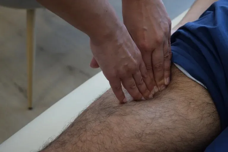
Assessment
Pain
Pain is one of the most used criteria in injury rehabilitation protocols. During each test, we must assess pain, as well as during strength and conditioning exercises. Some research has shown significant improvements after rehabilitation protocols using pain thresholds compared to pain-free rehabilitation
(Hickey et al., 2020). However, there is still a large amount of research recommending pain-free training. We must be cautious when training athletes with movement deficits or associated injuries like low back pain.
Several rehabilitation guides recommend tenderness to palpation as one of the criteria that we must control. Aspetar Hamstring Protocol recommends recording the length and width of the area of maximum pain and the distance between it and the ischial tuberosity.
In this first phase, we can record pain in daily activities such as walking, going up and down stairs, sitting and getting up, etc.
It is also essential to record pain in each test we perform.
Strength
The hamstrings, in addition to knee flexors and hip extensors, are knee stabilisers, especially during high-intensity running. As the rehabilitation progresses,
we will progress in speed, resistance, and range of motion. Isokinetic evaluation is the most precise and used method in research, but we can also use a dynamometer or manual resistance. The latter is the fastest and most effective assessment when we want to detect painful positions, although the other two forms will give us much more precise data at the end of the rehabilitation.
Knee flexion – The soccer player is lying prone for the first test, with the hip extended and the knee in 90º of flexion (Image 1). In this position, the soccer player performs three maximum isometric contractions of 3 seconds each, with a few seconds of rest between repetitions (If there is pain, we must stop). If there is no pain in this position, we can progress to the next test with the knee at about 30º of flexion and then in full extension. The progression continues with the player lying supine with the hip in 90º of flexion to evaluate the force with the knee at 90º, 45º, and maximum extension (Image 1).
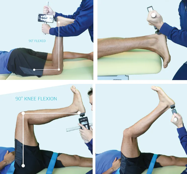
Image 1. Different positions to measure knee flexion strength
Hip extension – The glute bridge exercise may be used to assess hip extension ability. Although we will see how to evaluate the hip hinge pattern later, this exercise is also very useful and can help detect movement deficits. In this exercise, it is prevalent for players to exert more effort with the uninjured leg due to pain or fear after injury. If we detect this, I recommend evaluating the hip extension strength with the Prone Hip Extension Test (Image 2), placing the resistance in the popliteal region.

Image 2. Prone hip extension test (Schuermans, et al., 2017)
Lower limb stability – In several rehabilitation guides, evaluating the athlete’s stability and proprioception is also recommended. To begin, we can perform a Single Leg Squat to assess the stability of the lower limb and core and hip strength (Ugalde et al., 2015). If there is pain or insecurity, the test may be started by sitting on a high bench or a massage table, standing up with one leg, and returning to the starting position (Image 3).
In this test, we must evaluate if there is knee valgus or pelvic sway (Trendelenburg Sign) or if balance is lost due to instability of the upper limb.
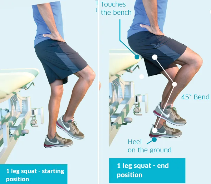
Image 3. Single leg Squat (Aspetar Hamstring Protocol)
If the footballer cannot perform a single leg Squat, the Trendelenburg Test may be used (Image 4 and Video 1). This test evaluates the stability of the pelvis and trunk during unilateral support (Wilshaw et al., 2020).
There is a modified version of the Trendelenburg Test that is arguably better suited for evaluating muscle function in athletes. In this test, the wall is not used as support, and the arms are free.
Deficits in trunk and pelvic control during the Modified Trendelenburg Test have been shown to be a risk factor in lower limb injuries
(Image 5), especially of the knee (Anterior cruciate ligament) (Leppänen et al., 2020).
The hamstrings play a crucial role in stabilising the knee. Deficits as a result of a hamstring injury, in combination with poor lumbopelvic control, can significantly increase the risk of suffering serious knee injuries.

Video 1. Trendelenburg Test
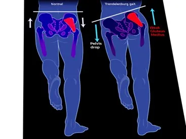
Image 4. Trendelenburg gait (Gandbhir, et al., 2021)
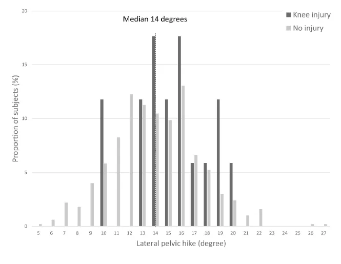
Image 5. Lateral pelvic hike during the standing knee lift test and proportion of subjects with knee injuries (Leppänen et al., 2020).
Range of Motion (ROM)
Hip flexion and knee extension ROM may be affected after a hamstring injury. I have chosen several tests based on different hip and knee positions to evaluate it. Each test can be performed actively or passively. In all tests, we must ensure no compensatory movement in the lumbopelvic area, especially at the end of the movement.
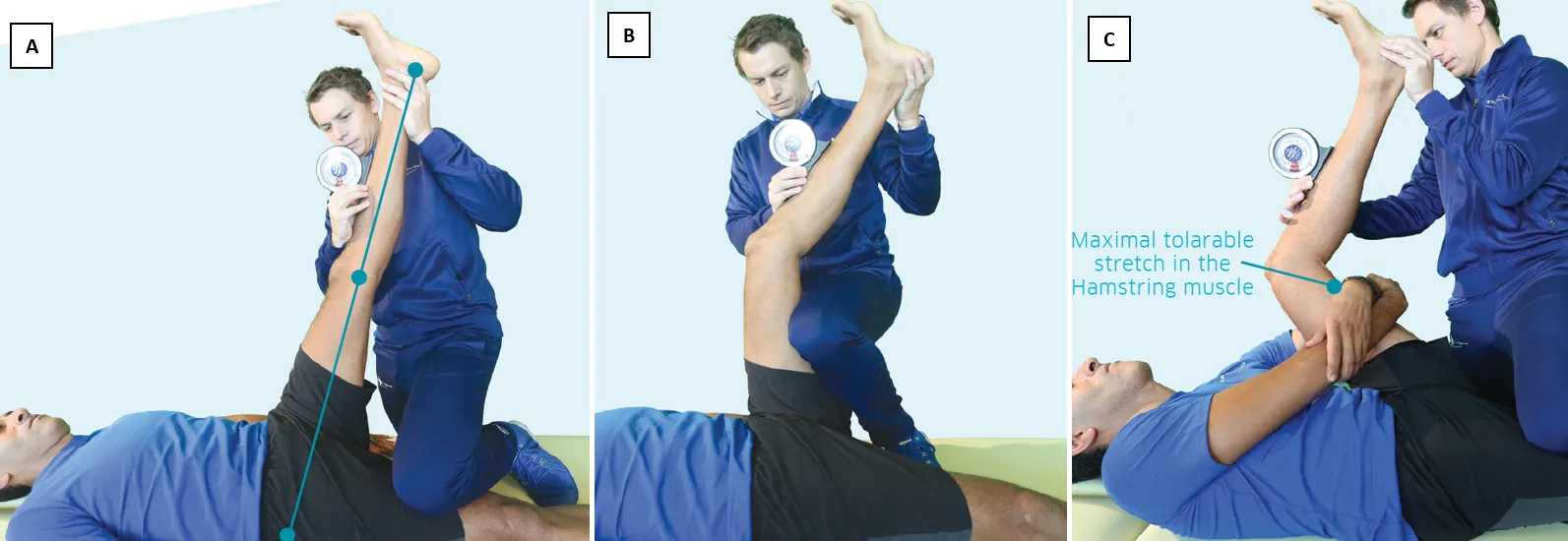
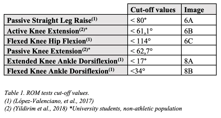
The Straight Leg Raise Test (Image 6A) is one of the most used tests to assess hip flexion ROM. With the patient lying supine, the leg is raised with the knee extended to the point where there is an onset of pelvic movement, or the patient feels a robust and tolerable stretch without pain (López-Valenciano et al., 2019).
The Knee Extension Test (Image 6B) also begins in a supine lying position, but now, the hip is flexed 90º, and the hands are held behind the knee. The maximum possible stretch is performed in this position following the same criteria as in the previous test (López-Valenciano et al., 2019). We can also evaluate knee extension ROM with the hip in maximum flexion as the Aspetar guide recommends. This test can also assess the maximum hip flexion angle prior to knee extension (Image 6 C).
Thomas Test (Image 7). In this test, the athlete lies supine with the hips on the table’s edge. With both arms, one leg is held in maximum hip flexion while the other is relaxed with the hip in extension. In this test, there are several movement patterns that can provide much information.
Restricted hip flexion (Image 7A) – This may be due to pain after an injury or muscle restrictions in the posterior musculature of the leg being held.
Restricted hip extension (Image 7B) –
The psoas is a muscle that tends to shorten and can generate anterior pelvic tilt. It is usually associated with excessive tension in the anterior thigh muscles. This shortening is also associated with gluteus maximus weakness due to reciprocal inhibition (Buckthorpe et al., 2019).
Restricted knee flexion (Image 7C) – A prevalent pattern in soccer players, big and powerful quadriceps, but shortened and under excessive tension.
Knee external rotation (Image 7D) –
One of the most common causes is excessive tension in the iliotibial band. The tensor fascia lata may be overactive due to weakness of the gluteal muscles (Selkowitz, et al., 2013).
This movement pattern is also associated with increased internal hip rotation and knee adduction, increasing the risk of knee injuries (Baker & Fredericson, 2016).

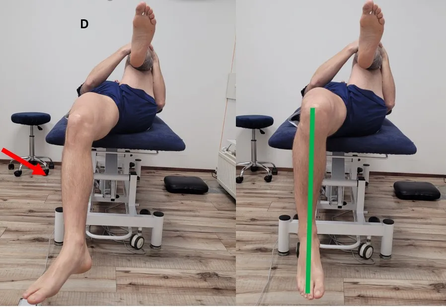
Image 7. Thomas Test
In professional soccer players, ankle mobility deficits have been observed (López-Valenciano et al., 2019). Research has shown that myofascial connections through the whole posterior muscle chain can transmit forces through the joints (Wilke et al., 2016), in this case, at the knee (Wilke et al., 2020). Although the relationship between hamstring injuries and mobility restrictions of the ankle-joint complex has not been researched, it may be interesting to investigate this further.
Although there are several tests to evaluate ankle mobility
(Mason-Mackay et al., 2017), weight-bearing variations seem to be most representative of the function performed by the lower limb during sports activities (Powden et al., 2015). In Image 8, two tests are shown, one with a flexed knee (Image 8A) and another with an extended knee (Image 8B).
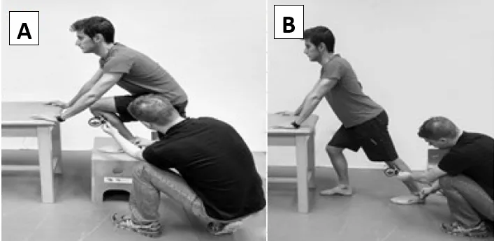
Image 8. Ankle mobility tests (López-Valenciano et al., 2019)
Table 1 shows reference values that have been used to evaluate hip, knee, and ankle ROM.
Lumbopelvic motor control
Pelvic control during high-intensity actions is crucial to the prevention of hamstring injuries. This is especially important for young football players.
In addition to specific motor patterns that require correct hip function, we must also evaluate lumbar stabilisation capacity when extension, flexion, rotation, and lateral flexion forces are exerted (Adelt et al., 2021).
The hip hinge is a fundamental movement pattern for the correct and effective execution of most exercises. If not performed well, the lumbar spine will compensate for the hip’s lack of mobility and strength. The Waiters Bow (Image 9A) is a test used to detect a lack of motor control during hip flexion.
This test evaluates the ability to flex the hip (50-70º) while standing in a neutral lumbar position (Luomajoki et al., 2007).
The test result will be negative if there is lumbar flexion or inability to reach at least 50º of hip flexion.
To evaluate control capacity over lateral flexion forces, we can use tests such as the modified Trendelenburg Test or the One Leg Stance Test (Meier et al., 2021) (Image 9B). The lateral displacement of the trunk in the last test should be less than 10 cm, and the difference between the right and left sides should be lower than 2 cm (Luomajoki et al., 2007).
Control over lumbar rotation can be evaluated with the crook lying test (Luomajoki et al., 2007) (Image 9C), in which abduction and external hip rotation are performed while lying supine with the knees flexed. There should be no compensatory movement of the pelvis or hip ROM deficits.
Lumbar hyperextension can cause excessive pelvic anterior tilt, increasing the risk of injury. For example, the Rocking Forwards Test (Image 9D) has been recommended to evaluate movement deficits in patients with low back pain (Meier et al., 2021). From the quadruped position, the hips are extended, and the body leans forward. As in all other tests, the result will be positive if the lumbar area is kept in a neutral position while the movement is performed.
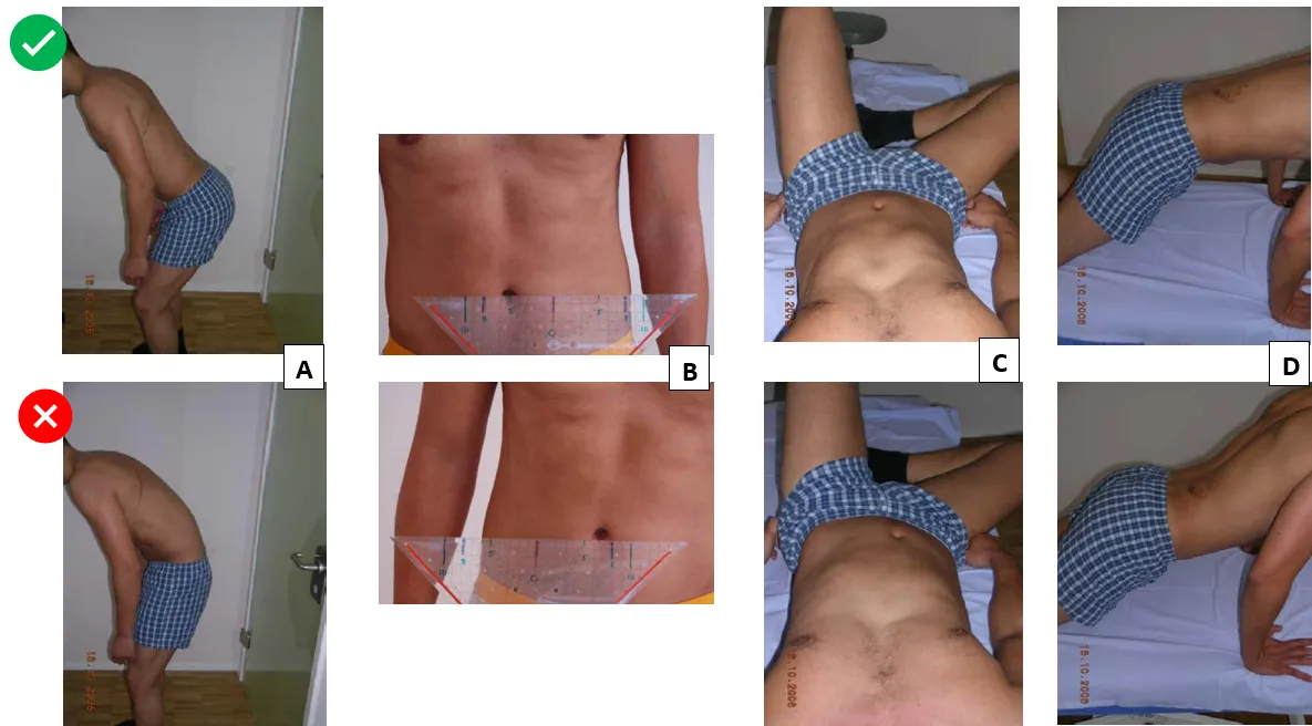
Image 9. Lumbopelvic assessment (Luomajoki et al., 2007); A: Waiters bow; B: One leg stance test; C: Crook lying test; D: Rocking forwards Test
For those of you who are interested in more lumbar stability assessment, I recommend the following literature: Adelt et al. (2021), Biele et al. (2019), and the work of Stuart McGill, especially his book Ultimate Back Fitness and Performance (2004).
