Ankle Dorsiflexion Mobility Assessment
VOETBAL MEDISCH SYMPOSIUM 2020
DE BEHANDELING VAN VOETBALBLESSURES
PRAKTISCHE WETENSCHAP
OP DE KNVB CAMPUS IN ZEIST VINDT KOMEND JAAR OPNIEUW HET VOETBALMEDISCH SYMPOSIUM PLAATS.
HET SYMPOSIUM IS DÉ PLEK OM COLLEGA’S BINNEN HET VOETBALMEDISCHE DOMEIN TE ONTMOETEN OF KENNIS OP TE DOEN VAN GERENOMMEERDE EXPERTS. EN DIE NIEUWSTE INNOVATIES TE ZIEN OP HET GEBIED VAN VOETBALMEDISCHE EN FYSIEKE PRESTATIES.
NA VORIG JAAR DE DIAGNOSTIEK VAN VOETBALBLESSURES BELICHT TE HEBBEN, ROLT DE BAL DIT JAAR VERDER NAAR DE BEHANDELING VAN VOETBALBLESSURES. HET INHOUDELIJKE PROGRAMMA BIEDT OPNIEUW SPREKERS DIE ZICH ONDERSCHEIDEN IN ZOWEL DE DAGELIJKSE ZORG VOOR DE VOETBALLERS ALS OP WETENSCHAPPELIJK GEBIED.
VOETBAL MEDISCHE WORKSHOP 2020
(VELD)REVALIDATIE NA EEN VOETBALBLESSURE
OP 4 MAART ZAL ER WEDEROM EEN WORKSHOP PLAATS VINDEN BIJ HET KNVB VOETBAL MEDISCH CENTRUM.
OOK DIT JAAR BELOOFD HET EEN OCHTENDVULLEND PROGRAMMA TE ZIJN WAAR VOORNAMELIJK (SPORT)FYSIOTHERAPEUTEN HUN KENNIS MEE KUNNEN UITBREIDEN.
TIJDENS DE WORKSHOP ZAL MATT TABERNER ZIJN KENNIS EN EXPERTICE MET DE DEELNEMERS GAAN DELEN. MATT TABERNER IS EEN ERVAREN CLINICUS DIE AL JAREN EINDVERANTWOORDELIJK IS VOOR DE REVALIDATIE VAN TOPVOETBALLERS IN DE PREMIER LEAGUE. ZIJN FOCUS LIGT VOORNAMELIJK OP FYSIEKE ONTWIKKELING EN PRESTATIES. TEVENS IS HIJ DE ONTWIKKELAAR VAN HET ‘CONTROL-CHAOS CONTINUUM’.
DIT FRAMEWORK, WELKE VIJF FASES BESCHRIJFT HOE DE VELDREVALIDATIE NA EEN VOETBALBLESSURE OPGEBOUWD KAN WORDEN, STAAT CENTRAAL BINNEN DE WORKSHOP. DE THEORETISCHE ACHTERGROND,
DE TOEPASSING EN HET PRAKTISCHE ASPECT ZULLEN ALLEN AAN BOD KOMEN TIJDENS DE WORKSHOP.
reviews

Nick van der Horst
Meet the soccerdoc
Nick van der Horst behaalde zijn diploma fysiotherapie in 2007 aan de Hogeschool Utrecht. Hij werkte 10 jaar lang als sportfysiotherapeut/echografist/docent bij het Academie Instituut te Utrecht. Daarna heeft hij de overstap gemaakt naar waar zijn hart ligt, het professionele voetbal. Hij heeft twee jaar als sportfysiotherapeut en hoofd van de medische staf bij Go Ahead Eagles in Deventer gewerkt. Momenteel is is Nick werkzaam bij de KNVB. Zijn onderzoeks-activiteiten zijn gefocust op de voetbal-medische zorg. In 2017 behaalde hij zijn doctoraal na het verdedigen van zijn proefschrift ‘Prevention of hamstring injuries in male soccer’.

Blogger: Raúl Gómez
The previous article demonstrated how restricted ankle dorsiflexion can impact landing mechanics by increasing ground reaction forces and causing medial knee displacement (Taylor et al., 2021). If you haven’t read it yet, we recommend that you do so in order to understand the importance of ensuring adequate ankle mobility in football players. Read here.
Football is a high-intensity sport with a high number of eccentric muscle actions that occur during changes of direction, jumps or decelerations. Continuous exposure to these types of movement may increase the tightness of the muscles and tendons and cause muscle damage, which will affect the neural properties of the myotendinous unit, reducing the total range of motion throughout the season (Moreno- Pérez et al., 2020), increasing the risk of injury (Mason-Mackay et al., 2017) and negatively influencing sports performance (Gonzalo-Skok et al., 2015).
Although there is general agreement that the lack of ankle dorsiflexion increases the risk of lower limb injuries (Mason-Mackay et al., 2017), there is a notable difference in the evaluations utilized in various scientific articles and in the cut-off values for each test. In addition, there is no consensus on the optimal amount of mobility of this joint and how (lack of) mobility is associated with injury risk.
The tests used to assess ankle mobility can be classified into three categories:
- Open Kinetic Chain (OCC)
- Closed Kinetic Chain (CCC)
- Landing mechanics
Different methods have been used to measure the results of each test, such as a goniometer, inclinometer, or analysis of digital models. In this article, we will not discuss the measurement method.
Open Kinetic Chain (OKC)
This type of evaluation is the least functional to sports activity since all the actions where ankle mobility is important are performed in closed kinetic chains (landings, changes of direction, running, etc.). Although this type of test will not provide us with any information about the control and mobility that the football player has in specific actions, it can be very useful when evaluating mobility restrictions or pain on specific structures.
In these tests, ankle dorsiflexion is evaluated with the patient sitting or lying on the treatment table. The ankle is passively dorsiflexed until the limit of movement is reached. This test should be performed first with the knee extended to assess flexibility of the gastrocnemius muscle, and then with the knee flexed at 90º to assess flexibility of the soleus. (Stovitz & Coetzee, 2004). Stovitz & Coetzee (2004) recommend testing this musculature with the subtalar joint in a neutral position while lateral force is applied to the neck of the talus and the forefoot is pushed medially (Image 1).
In this way, the foot is locked, preventing the eversion of the calcaneus, which could give a false impression of greater flexibility. Ankle dorsiflexion can be limited not only by restrictions in muscle flexibility, but also by intra-articular factors such as tightness of the talocrural posterior capsule, positional faults of the fibula, or reduced posterior glide of the talus relative to the ankle mortise (Howe, 2020).
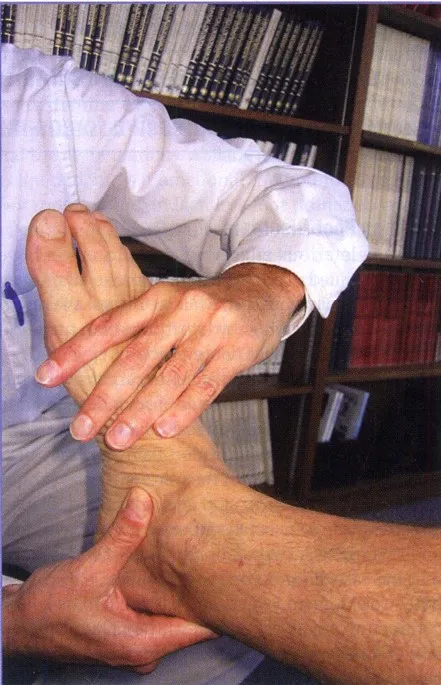
Image 1. OKC ankle mobility assessment. (Stovitz & Coetzee, 2004)
Several authors recommend that the range of motion in these tests should reach 20º of dorsal flexion (Brockett & Chapman, 2016) (Magee, 2014). Stiffler et al. (2014), compared range of motion values during this type of test in individuals who do not show medial knee displacement during an overhead squat compared to those who do (Image 2). Individuals with greater ankle dorsiflexion range of motion showed less medial knee displacement.
Image 2. Range of motion in OKC tests in the study by Stiffler et al. (2014); MKD – Medial knee displacemen
Due to the significant variation among studies, it is challenging to propose cut-off values. It is essential to compare both legs and ensure the absence of unilateral deficits. There is not much reference data on this type of evaluation in soccer players.
Closed Kinetic Chain (CKC)
In comparison to OKC tests, CKC assessments provide more information about ankle joint mobility in positions similar to those that occur during sports activities. Movement deficits during this type of test have been related to movement alterations that increase the risk of injury, such as knee valgus, excessive hip adduction, or greater trunk inclination (Ivana Hanzlíková et al., 2022).
There are many variations of CKC assessment. One test that is frequently applied is the Weight Bearing Lunge Test (WBLT) or Wall Test (Image 3). In this test, the subject is placed in a lunge position facing a wall, with one leg in front of the other. From this position, the patient lunges forward until the knee touches the wall, keeping the heel on the floor. If there is contact with the wall, the foot is moved back until finding the farthest distance where the knee contacts the wall while the heel remains entirely in contact with the ground. When the limit of movement is reached, the therapist measures the distance from the wall to the tip of the big toe.
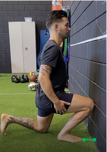
Image 3. Weight Bearing Lunge Test or Wall test
There are variations of this test, such as the Leg Motion System Test (Moreno-Pérez et al., 2020) or the Half Kneeling Dorsiflexion Test (Gourlay et al., 2019), but they all consist of the same movement, only varying the measuring device or the position of the rear leg.
Dill et al. (2014) suggested a cutoff value of 44.02º for this test, while other authors, such as Moreno-Pérez et al. (2020), did not specify cutoff values in their studies but indicated that differences of more than 2 cm between legs should be considered as asymmetries that increase the risk of injury. Once again, it is challenging to establish exact cutoff values for soccer players due to the wide range of results in different studies.
The Modified Weight-Bearing Lunge Test, also known as the Ankle Clearing Test (Image 4), is performed with the patient standing with one leg in front of the other. The heel of the front leg is in contact with the toes of the back leg. A dowel is given to the patient to help maintain balance. From this position, the patient moves the knee of the rear leg forward as far as possible, keeping the heel in contact with the ground.
The therapist measures the position of the knee in relation to the medial malleolus of the ankle of the front leg: behind, in line, or beyond (Gourlay, et al., 2019). This test has been shown to be a valid tool in evaluating ankle dorsiflexion and has been compared with the values obtained in the WBLT.
The modified WBLT score corresponds to 33.5 ± 2.0 degrees if the knee is behind the medial malleolus of the front leg, 38.6 ± 1.2 degrees if it is in line, and 43.0 ± 0.78 degrees if it is beyond (Gourlay, et al., 2019). Therefore, soccer players with adequate ankle mobility should be able to move the knee beyond the medial malleolus of the front leg.
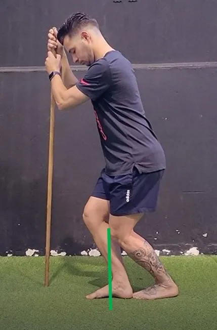
Image 4.
Modified Weight Bearing Lunge Test or Ankle clearing test
Image 5 shows two other ankle dorsiflexion tests. One with the knee in extension (Right, the gastrocnemius will limit the result of this test to a greater extent) and another with the knee in flexion (Left, the soleus will limit the result of this test to a greater extent).
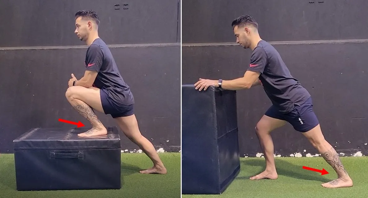
Image 5.
Tests variations for the measurement of ankle dorsiflexion. Knee in flexion (Front leg; Left); Knee Extension (Back Leg; Right)
López-Valenciano et al. (2019), in a study with professional soccer players, used cut-off values of <17º for the test with the knee in extension and &<34º for the test with the knee in flexion. Lower values in these tests would be considered movement restrictions, increasing the risk of lower limb injuries.
Landing Mechanics
Various types of landing mechanics assessment have been used to assess lower limb movement mechanics. To summarize, we propose three groups: Bilateral, unilateral, and jump after landing. Once again, due to the wide variety in methodology (such as jump height, measurement method, and instructions during landing), it is challenging to propose standard tests and cut-off values.
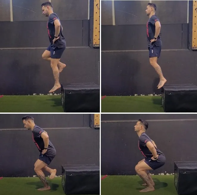
Image 6. Unilateral and bilateral landing
One of the most used tests in research is the Landing Error Scoring System” (LESS) test. It is performed from a 30 cm high box, from which the subject jumps forward at a distance equivalent to 50% of the subject’s height. In this test, the landing is bilateral. The system utilizes a 17-item scoring method to identify athletes who are at risk of injury. It is used to assess the ankle and the entire lower limb. In a study conducted by Padua et al. (2015), an example is provided to demonstrate how this evaluation method can be applied to soccer players. You can find the details in the references.
Whitting et al. (2011), conducted a study based on single-leg drop landings from heights of 32 and 72 cm. Malloy et al. (2014) assessed landing mechanics by having participants perform a maximum vertical jump immediately after landing from a box jump, with the height of the box matching the maximum height reached in a previous vertical jump by each participant. In both studies, researchers observed altered movement patterns in participants with limited ankle mobility measured by static tests.
Conclusion
OKC evaluations can help us detect deficits in isolated structures in the early stages of rehabilitation. However, once we have corrected the deficits, we must progress to tests and exercises with more functionality (e.g. CKC tests) and ensure the player is ready for competition.
The CKC tests are a quick and effective way to assess ankle mobility and identify soccer players at higher risk of injury. This makes them a good option when planning injury prevention programs. However, there is debate about whether static assessments are useful in predicting injuries. Even if a football player has good ankle mobility, we don’t know if they use an appropriate landing strategy. There may also be other deficits that predispose the football player to a higher risk of injury, such as excessive knee valgus due to weakness of the gluteal muscles, lack of lumbopelvic control or poor ankle stiffness.
Evaluating the landing mechanics of soccer players who have suffered long-term injuries, such as to the anterior cruciate ligament, or who experience chronic pain, for example, in the Achilles tendon or patellofemoral area, can provide essential information before they finish their rehabilitation and return to train or match play.
In our upcoming blog, the last of this series, we will explore different methods to enhance ankle mobility. We will identify which structures limit the movement and which ones are weakened or inhibited and propose targeted mobilizations and exercises to improve ankle dorsiflexion.
References
Brockett, C., & Chapman, G. (2016). Biomechanics of the ankle. Orthopaedics and trauma, 30(3), 232-238.
Dill, K., Begalle, R., Frank, B., Zinder, S., & Padua, D. (2014). Altered Knee and Ankle Kinematics During Squatting in Those With Limited Weight-Bearing–Lunge Ankle Dorsiflexion Range of Motion. Journal of Athletic Training, 49(6), 723-732.
Gonzalo-Skok, O., Serna, J., Rhea, M. R., & Marin, P. J. (2015). Relationships between Functional Movement Tests and Performance Tests in Young Elite Male Basketball Players. International Journal of Sports Physical Therapy, 10(5), 628-638.
Gourlay, J., Bullock, G., Weaver, A., Matsel, K., Kiesel, K., & Plisky, P. (2019). The Reliability and Criterion Validity of a Novel Dorsiflexion Range of Motion Screen. Athletic Training and Sports Health Care, 12(1).
Howe, L. P. (2020). Restrictions in ankle dorsiflexion range of motion and its effect on landing mechanics. Thesis, Ede Hill University.
Ivana Hanzlíková, I., Richards, J., & Hébert Losier, K. (2022). The influence of ankle dorsiflexion range of motion on unanticipated cutting kinematics. Sport Sciences for Health.
López-Valenciano, A., Ayala, F., Vera-García, F., De Ste Croix, M., Hernández, S., Ruiz, I., Cejudo, A., Santonja, F. (2019). Comprehensive profile of hip, knee and ankle ranges of motion in professional football players. The Journal of Sports Medicine and Physical Fitness, 59(1), 102-109.
Magee, D. (2014). Orthopedic Physical Assessment (Sixth ed.). St Louis, Missouri: Elsevier.
Malloy, P., Meinerz, C., Geiser, C., & Kipp, K. (2015). The Association of Dorsiflexion Flexibility on Landing Mechanics during a Drop Vertical Jump. Knee Surgery, Sports Traumatology, Arthroscopy, 23(12).
Mason-Mackay, A., Whatman, C., & Reid, D. (2017). The effect of reduced ankle dorsiflexion on lower extremity mechanics during landing: A systematic review. Journal of Science and Medicine in Sport, 20, 451–458.
Moreno-Pérez, V., Soler, A., Ansa, A., López-Samanes, Á., Madruga-Parera, M., Beato, M., & Romero-Rodríguez, D. (2020). Acute and chronic effects of competition on ankle dorsiflexion ROM in professional football players. European Journal of Sports Science, 20(1), 51-60.
Padua, D., DiStefano, L., Beutler, A., de la Motte, S., DiStefano, M., & Marshall, S. (2015). The Landing Error Scoring System as a Screening Tool for an Anterior Cruciate Ligament Injury–Prevention Program in Elite-Youth Soccer Athletes. Journal of Athletic Training, 50(6), 589-595.
Stiffler, M., Pennuto, A., Smith, M., Olson, M., & Bell, D. (2014). Range of Motion, Postural Alignment, and LESS Score Differences of Those With and Without Excessive Medial Knee Displacement. Clinical journal of sport medicine: official journal of the Canadian Academy of Sport Medicine, 25, 61-66.
Stovitz, S., & Coetzee, C. (2004). Hyperpronation and Foot Pain – Steps Toward Pain-Free Feet. The Physician and Sportsmedicine, 32(8), 19-26.
Taylor, J., Wright, E., Waxman, J., Schmitz, R., Groves, J., & Shultz, S. (2021). Ankle dorsiflexion affects hip and knee biomechanics during landing. Sports health.
Whitting, J., Steele, J., McGhee, D., & Munro, B. (2011). Dorsiflexion Capacity Affects Achilles Tendon Loading during Drop Landings. MEDICINE & SCIENCE IN SPORTS & EXERCISE, 43(4), 706-713.

